. 2012 Jun;86(12):6932-46.
doi: 10.1128/JVI.07147-11. Epub 2012 Apr 11.

Mice deficient in STAT1 but not STAT2 or IRF9 develop a lethal CD4+ T-cell-mediated disease following infection with lymphocytic choriomeningitis virus

Affiliations

Mice deficient in STAT1 but not STAT2 or IRF9 develop a lethal CD4+ T-cell-mediated disease following infection with lymphocytic choriomeningitis virus

Markus J Hofer et al. J Virol. 2012 Jun.

Abstract

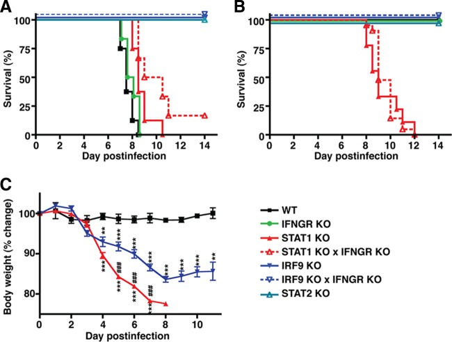
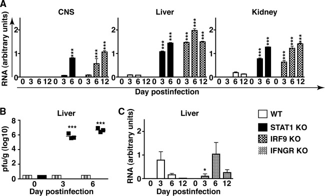
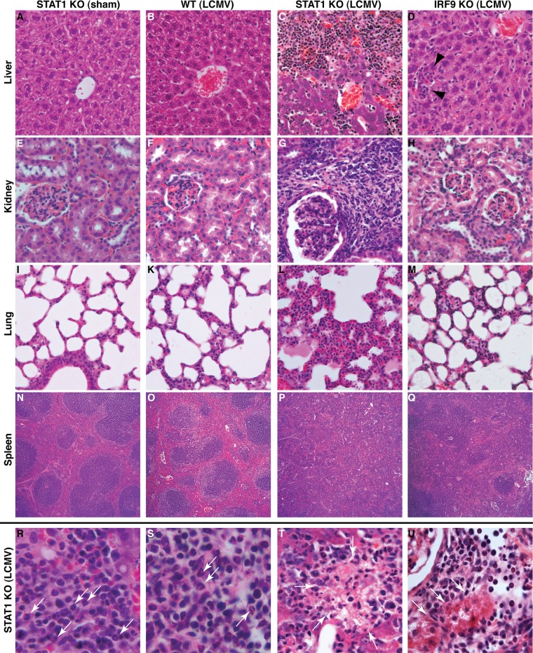
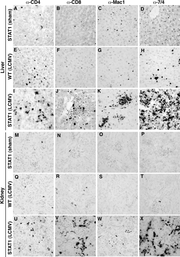
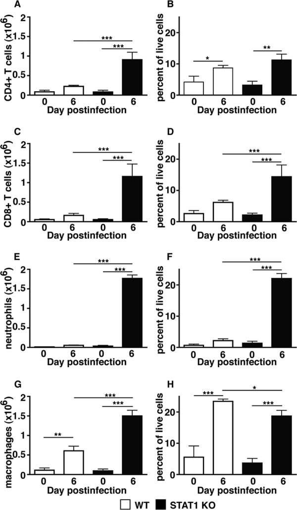
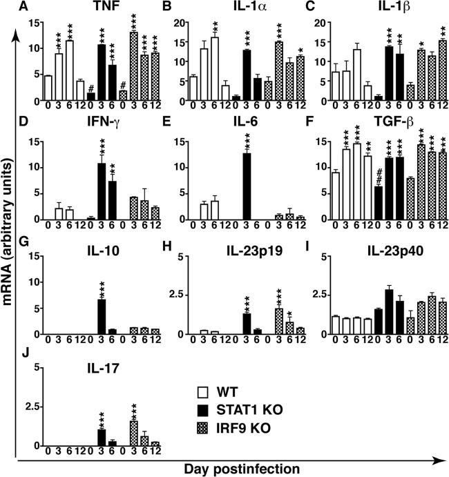
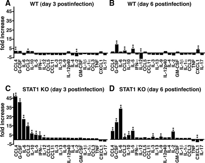
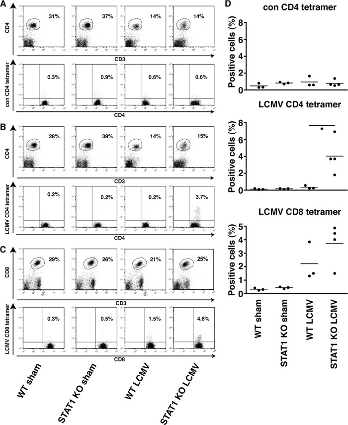
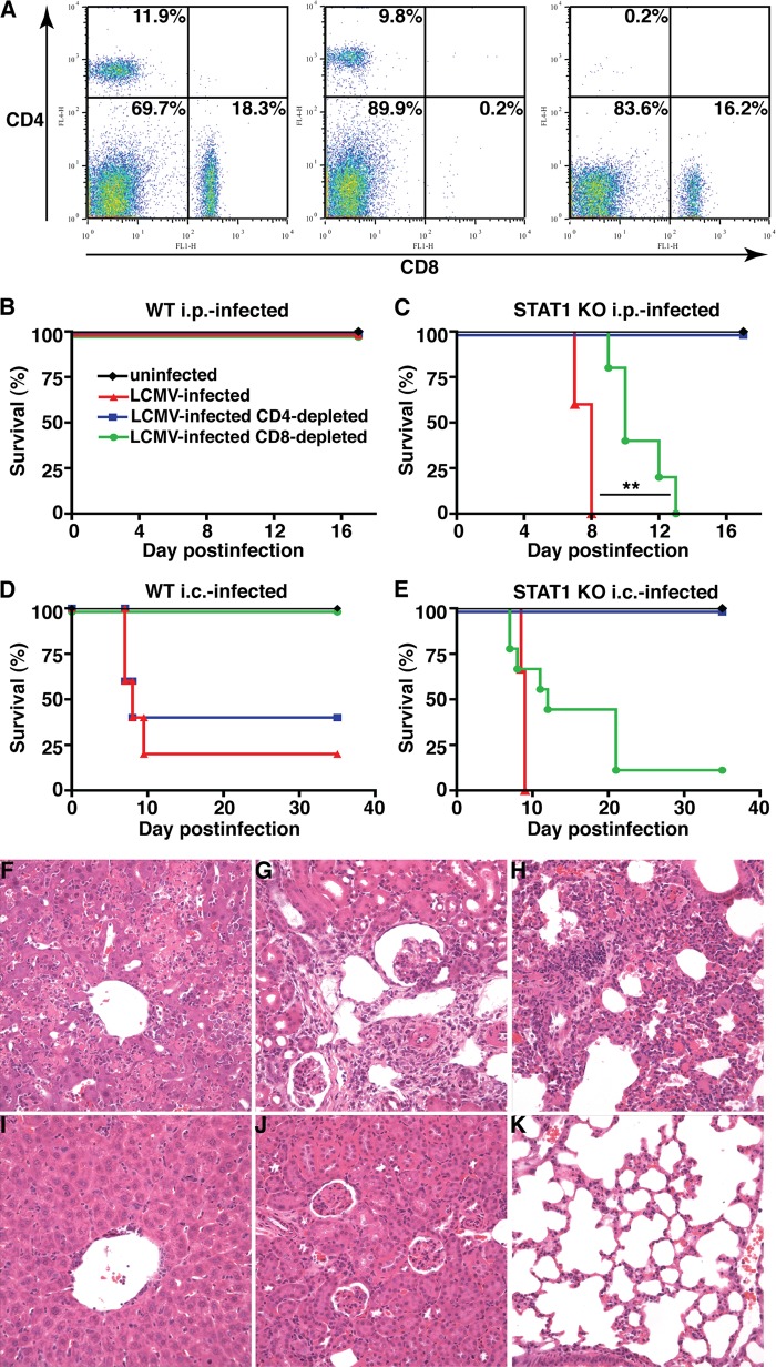
Interferon (IFN) signaling is crucial for antiviral immunity. While type I IFN signaling is mediated by STAT1, STAT2, and IRF9, type II IFN signaling requires only STAT1. Here, we studied the roles of these signaling factors in the host response to systemic infection with lymphocytic choriomeningitis virus (LCMV). In wild-type (WT) mice and mice lacking either STAT2 or IRF9, LCMV infection was nonlethal, and the virus either was cleared (WT) or established persistence (STAT2 knockout [KO] and IRF9 KO). However, in the case of STAT1 KO mice, LCMV infection was lethal and accompanied by severe multiorgan immune pathology, elevated expression of various cytokine genes in tissues, and cytokines in the serum. This lethal phenotype was unaltered by the coabsence of the gamma interferon (IFN-γ) receptor and hence was not dependent on IFN-γ. Equally, the disease was not due to a combined defect in type I and type II IFN signaling, as IRF9 KO mice lacking the IFN-γ receptor survived infection with LCMV. Clearance of LCMV is mediated normally by CD8(+) T cells. However, the depletion of these cells in LCMV-infected STAT1 KO mice was delayed, but did not prevent, lethality. In contrast, depletion of CD4(+) T cells prevented lethality in LCMV-infected STAT1 KO mice and was associated with a reduction in tissue immune pathology. These studies highlight a fundamental difference in the role of STAT1 versus STAT2 and IRF9. While all three factors are required to limit viral replication and spread, only STAT1 has the unique function of preventing the emergence of a lethal antiviral CD4(+) T-cell response.


Figures

Fig 1
Fig 1
STAT1 deficiency causes a lethal disease in mice infected with LCMV. (A) I.c. infection of WT and IFNGR-deficient mice with LCMV caused LCM with characteristic seizures and death. STAT2 KO and IRF9 KO mice developed transient, mild clinical signs following i.c. infection and survived. In contrast, i.c.-infected STAT1 KO mice developed a progressive lethal wasting disease but no seizures. (B) I.p. infection of WT, IFNGR KO, STAT2 KO, and IRF9 KO mice resulted in mild clinical signs, and none of the mice died. Comparable with i.c. infection, i.p. infection of STAT1 KO mice caused a wasting disease and the death of all animals. (C) In contrast to WT mice, starting at day 4 postinfection, i.p. infection with LCMV resulted in a significant reduction of the body weight, by approximately 10 to 15% in IRF9 KO mice and more than 20% in STAT1 KO mice, by day 7 postinfection. Loss of body weight was significantly greater in STAT1 KO mice than in IRF9 KO mice, and in the latter, body weight stabilized from day 8 postinfection. The data are shown as means ± standard errors of the mean (SEM). ** and ***, P < 0.01 and P < 0.001 compared with WT mice; ###, P < 0.001 compared with IRF9 KO mice, as determined by Mann-Whitney U test.
Fig 2
Fig 2
STAT1 and IRF9 are required to limit viral replication and spread and for the elimination of LCMV. (A) WT mice infected i.p. had low levels of LCMV-NP RNA in the liver at days 3 and 6 postinfection and in the kidney at days 6 and 12 postinfection. No viral RNA was detectable in the central nervous system (CNS). High levels of viral RNA were present in the livers, kidneys, and CNS of infected STAT1 KO and IRF9 KO mice. (B) Plaque assays performed on liver lysates from WT and STAT1 KO mice showed no infectious virus detectable by the assay in uninfected WT and STAT1 KO mice or in infected WT mice. In contrast, a high virus titer was present in the livers of day 3 and day 6 infected STAT1 KO mice. (C) Similar to WT mice, LCMV-NP RNA was detectable in the liver, but not the CNS, of IFNGR KO mice. Viral clearance was delayed in IFNGR mice compared with WT mice. Values from RPA were normalized to the housekeeping gene L32 and are shown as mean and SEM. *, P < 0.05, and ***, P < 0.001 compared with the respective time point in WT mice as determined by Mann-Whitney U test.
Fig 3
Fig 3
LCMV infection of STAT1 KO mice results in destructive organ pathology. The livers (A), kidneys (E), and lungs (I) of sham-infected STAT1 KO mice showed no overt pathological alterations, and germinal centers present in the spleen appeared normal (N). Comparable findings were obtained for the livers (B), kidneys (F), lungs (K), and spleens (O) from WT mice at day 6 postinfection. In contrast, the livers (C), kidneys (G), and lungs (L) of infected STAT-deficient mice showed prominent mononuclear infiltrates. No organized germinal centers were seen in the spleens (P) of infected STAT1 KO mice. The livers (D, arrowheads), kidneys (H), and lungs (M) from infected IRF9 KO mice contained few foci with infiltrating leukocytes. In the spleens (Q) of infected IRF9 KO mice, germinal centers were less easily discernible than in WT mice but were still identifiable. Higher magnification of tissue sections from the livers (R) and kidneys (S) of infected STAT1 KO mice showed the presence of lymphocytes and polymorphonuclear granulocytes (arrows), as well as focal necrosis, in liver (T, arrows) and kidney (U, arrows; note the normal glomerulus in the top left corner). Original magnifications: A to H, ×20; I to M, ×10; N to Q, ×5; R to U, ×40.
Fig 4
Fig 4
Immunophenotype of infiltrating leukocytes in the livers and kidneys of STAT1 KO mice infected with LCMV. In sham-infected STAT1 KO mice, low numbers of CD4+ T cells (A and M), CD8+ T cells (B and N), macrophages (C and O), and neutrophils (D and P) were present in the liver and kidney, respectively. At day 6 post-i.p. infection, few infiltrating CD4+ (E and Q) and CD8+ (F and R) T cells, macrophages (G and S), and neutrophils (H and T) were seen in the livers and kidneys of WT mice. In contrast, numerous CD4+ (I and U) and CD8+ (J and V) T cells, macrophages (K and W), and neutrophils (L and X) infiltrated the livers and kidneys of infected STAT1 KO mice at day 6 postinfection. Original magnification (all panels), ×20.
Fig 5
Fig 5
Significantly increased numbers of leukocytes infiltrate the livers of LCMV-infected STAT1 KO mice. Shown is FACS analysis of leukocytes isolated from the livers of WT and STAT1 KO mice at day 6 postinfection. Compared with uninfected WT mice, livers from infected WT mice showed slight increases in the numbers of CD4+ (A) and CD8+ (C) T cells, neutrophils (E), and macrophages (G). In contrast, large numbers of CD4+ (A) and CD8+ (C) T cells, neutrophils (E), and macrophages (G) infiltrated the liver in STAT1 KO mice following infection. Notably, neutrophils were the most frequent cell type, followed by macrophages and CD8+ T cells. (B, D, F, and H) Percentages of populations in respect to all live cells. Data are shown as means and SEM. *, **, and ***, P < 0.05, P < 0.01, and P < 0.001 as determined by Mann-Whitney U test.
Fig 6
Fig 6
Upregulation of various proinflammatory and counterinflammatory cytokine genes in the livers of LCMV-infected WT and STAT1 KO mice. (A to J) Results of densitometric quantification of RPA autoradiographs. The values were normalized to the housekeeping gene L32 and are shown as means and SEM. *, P < 0.05; ** and ##, P < 0.01; *** and ###, P < 0.001 compared with the uninfected control of the same genotype (*) or WT mice (#) as determined by one-way analysis of variance (ANOVA).
Fig 7
Fig 7
Several cytokines are increased significantly in the sera of infected STAT1 KO mice. In WT mice, a multiplex assay showed small to moderate but significant changes for CCL4, CCL2, and IL-2 levels at day 3 postinfection (A) and for G-CSF, CCL2, IL-5, IL-12, and CXCL1 levels at day 6 postinfection (B). In contrast, at day 3 postinfection (C), the sera of STAT1 KO mice contained significantly higher levels of CCL4, G-CSF, IL-6, CCL2, IL-10, IL-5, IFN-γ, and IL-12 p70 than those of sham-infected controls. (D) At day 6 postinfection, CCL4, G-CSF, IL-6, CCL2, IL-10, IL-5, IL-3, IL-9, and IL-2 were significantly elevated in the sera of LCMV-infected STAT1 KO mice compared with sham-infected mice. In STAT1 KO mice, CXCL1 levels were significantly lower at day 6 postinfection than in the sham-infected controls. *, P < 0.05; **, P < 0.01 compared with the uninfected control as determined by one-way ANOVA.
Fig 8
Fig 8
Increased percentages of virus-specific CD4+ and CD8+ T cells in LCMV-infected STAT1 KO mice. (A and D) Representative FACS results from lymph node cells stained with CD3, CD4, and control MHC class II tetramer showed no difference between WT and STAT1 KO mice sham injected or infected with LCMV. (B and D) There was no significant increase in CD4+ T cells from infected WT mice compared with uninfected WT mice that recognized LCMV GP 66-77 MHC class II. In contrast, significantly more CD4+ T cells from infected STAT1 KO mice than from uninfected STAT1 KO mice bound the LCMV GP66-77 MHC class II tetramer. (C and D) Similarly, significantly more CD8+ T cells from infected STAT1 KO mice bound the LCMV GP33-41 MHC class I than uninfected mice, whereas there was no difference between infected and uninfected WT mice. *, P < 0.05 as determined using the one-tailed Mann-Whitney U test.
Fig 9
Fig 9
CD4+ T cells are required for lethal disease in LCMV-infected STAT1 KO mice. (A) Compared with Ig-treated controls (left), mice administered neutralizing monoclonal antibodies against CD8 (middle) or CD4 (right) showed less than 0.5% remaining CD8+ and CD4+ cells, respectively. Depletion of CD4+ T cells had no impact on the survival of WT mice infected i.p. or i.c. with LCMV (B and D) but prevented lethal disease in STAT1 KO mice independent of the route of infection (C and E). In contrast, depletion of CD8+ T cells rescued WT mice infected i.c. with LCMV (D) but did not alter the clinical course of WT mice infected i.p (B) or of STAT1 KO mice infected i.c. (E) with LCMV. (C) Depletion of CD8+ T cells significantly (**, P < 0.05) increased the survival time of STAT1 KO mice infected i.p. with LCMV but did not prevent lethal disease. Prominent mononuclear infiltrates and tissue destruction were evident in the livers (F), kidneys (G), and lungs (H) of STAT1 KO mice infected i.p. with LCMV. In contrast, in similarly infected STAT1 KO mice that were depleted of CD4+ T cells, the livers (I), kidneys (J), and lungs (K) showed only minor mononuclear infiltrates and no obvious organ destruction.

Similar articles

Cited by

References

    1. Ahmed R, Butler LD, Bhatti L. 1988. T4+ T helper cell function in vivo: differential requirement for induction of antiviral cytotoxic T-cell and antibody responses. J. Virol. 62:2102–2106 - PMC - PubMed
    1. Ahmed R, King CC, Oldstone MB. 1987. Virus-lymphocyte interaction: T cells of the helper subset are infected with lymphocytic choriomeningitis virus during persistent infection in vivo. J. Virol. 61:1571–1576 - PMC - PubMed
    1. Aichele P, et al. 2006. CD8 T cells specific for lymphocytic choriomeningitis virus require type I IFN receptor for clonal expansion. J. Immunol. 176:4525–4529 - PubMed
    1. Allan JE, Doherty PC. 1990. Binding of monoclonal antibodies and T cell effector function in vivo. Hybridoma 9:9–15 - PubMed
    1. Alsharifi M, Mullbacher A, Regner M. 2008. Interferon type I responses in primary and secondary infections. Immunol. Cell Biol. 86:239–245 - PubMed

Publication types

MeSH terms

Substances