. 2012 Jun 8;287(24):20522-33.
doi: 10.1074/jbc.M111.323279. Epub 2012 Apr 10.

Paired helical filaments from Alzheimer disease brain induce intracellular accumulation of Tau protein in aggresomes

Affiliations

Paired helical filaments from Alzheimer disease brain induce intracellular accumulation of Tau protein in aggresomes

Ismael Santa-Maria et al. J Biol Chem. .

Abstract

Abnormal folding of tau protein leads to the generation of paired helical filaments (PHFs) and neurofibrillary tangles, a key neuropathological feature in Alzheimer disease and tauopathies. A specific anatomical pattern of pathological changes developing in the brain suggests that once tau pathology is initiated it propagates between neighboring neuronal cells, possibly spreading along the axonal network. We studied whether PHFs released from degenerating neurons could be taken up by surrounding cells and promote spreading of tau pathology. Neuronal and non-neuronal cells overexpressing green fluorescent protein-tagged tau (GFP-Tau) were treated with isolated fractions of human Alzheimer disease-derived PHFs for 24 h. We found that cells internalized PHFs through an endocytic mechanism and developed intracellular GFP-Tau aggregates with attributes of aggresomes. This was particularly evident by the perinuclear localization of aggregates and redistribution of the vimentin intermediate filament network and retrograde motor protein dynein. Furthermore, the content of Sarkosyl-insoluble tau, a measure of abnormal tau aggregation, increased 3-fold in PHF-treated cells. An exosome-related mechanism did not appear to be involved in the release of GFP-Tau from untreated cells. The evidence that cells can internalize PHFs, leading to formation of aggresome-like bodies, opens new therapeutic avenues to prevent propagation and spreading of tau pathology.


Figures

FIGURE 1.
FIGURE 1.
Internalization of PHFs by fluid-phase endocytosis. A, unlabeled PHFs stained with uranyl acetate and examined by electron microscopy and PHFs labeled with Cy5 (46) and assessed by Western blotting and 10-nm-immunogold electron microscopy (inset) using anti-Cy5 antibody. B and C, HEK 293T cells treated with PHF-Cy5 alone (B) or HEK 293T cells treated with both PHF-Cy5 and Texas Red-dextran (C) for 6 h and stained with cholera toxin subunit B (CT-B)-Alexa Fluor 488 conjugate to label lipid rafts in the plasma membrane. Cell nuclei were stained with DAPI. Arrows indicate internalized PHF-Cy5 aggregates (in B) and their co-localization with Texas Red-dextran (in C). An asterisk denotes an extracellular PHF-Cy5 aggregate. Orthogonal projections in merged panels confirm internalization and co-localization of PHF-Cy5 aggregates. Imaging was performed using a scanning confocal microscope, LSM 700 (Zeiss).
FIGURE 2.
FIGURE 2.
Exogenous PHFs induce intracellular aggregation of tau in HEK 293T cells. A, untreated cells expressing GFP-Tau. B, PHF-treated cells showing GFP-Tau-positive aggregates (arrowheads). C, lactacystin-treated cells showing GFP-Tau-positive aggregates (arrowheads). D, GFP-expressing cells treated with lactacystin showing GFP-positive inclusions (arrowhead). E, quantitation of cells containing perinuclear aggregates. Data are means ± S.E.; n = 6 (n = 5 for GFP + PHFs). n.s., p > 0.05; *, p < 0.05; and ***, p < 0.001 as compared with untreated controls (None) using one-way analysis of variance and a post hoc Tukey's test for multiple comparisons among the six groups.
FIGURE 3.
FIGURE 3.
Sarkosyl-soluble and -insoluble tau content with or without extracellular PHFs. A, Western blotting of Sarkosyl-soluble and -insoluble fractions from SH-SY5Y cells with or without expression of GFP-Tau treated or untreated with PHFs. Gels are 10% polyacrylamide. B, densitometric quantitation of Sarkosyl-soluble and -insoluble fractions from HEK 293T cells expressing or not expressing GFP-Tau and treated or untreated with PHFs. Antibody 7.51 against total tau was used. Data are means ± S.D.; n = 3. *, p < 0.05 as compared with untreated controls by Student's t test. Lenti, lentivirus.
FIGURE 4.
FIGURE 4.
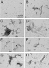
Immunogold electron microscopy of Sarkosyl-insoluble tau aggregates. Sarkosyl-insoluble fractions from untreated HEK 293T cells (A) and HEK 293T cells treated with AD PHFs (B–H) for 24 h and labeled with immunogold particles. A–E, anti-GFP (10-nm gold); F, PHF-1 for phosphorylated tau (25-nm gold); G, total human tau (HT7; 25-nm gold); and H, Ser(P)214tau (10-nm gold). A Hitachi H7000 electron microscope was used.
FIGURE 5.
FIGURE 5.
PHFs cause formation of aggresomes co-localized with γ-tubulin. Immunocytochemistry of SH-SY5Y cells expressing GFP-Tau (green) and either not treated (No PHFs) or treated with PHFs (+AD PHFs) for 24 h is shown. Cells were fixed and stained for γ-tubulin (Alexa Fluor 594; red). Arrows show perinuclear region without (A–C) or with aggregated GFP-Tau-positive inclusions (D–F) co-localized with the γ-tubulin-positive centrosome.
FIGURE 6.
FIGURE 6.
PHFs cause changes in vimentin filament network. Immunocytochemistry of SH-SY5Y cells expressing GFP-Tau (green) and either not treated (No PHFs) or treated with PHFs (+AD PHFs) for 24 h. Cells were fixed and stained for vimentin (Alexa Fluor 594; red). GFP-Tau-positive perinuclear inclusions are marked with asterisks. Redistributed vimentin filaments are marked with arrows.
FIGURE 7.
FIGURE 7.
Exosomes secreted by SH-SY5Y cells do not contain GFP-Tau. Total cell homogenates (Cell) and exosomes (Exosome) isolated from the conditioned medium of control (Ctrl) and GFP-Tau-overexpressing (GFP-Tau) SH-SY5Y cells and unconditioned medium (UM) (see “Experimental Procedures”). A, staining for flotillin as an exosomal marker protein. B, anti-tau clone RD3 binding three-repeat tau at ∼55 kDa. C, anti-tau clone RD4 binding four-repeat tau at ∼48 and ∼55 kDa and the GFP-Tau construct (*) at 90 kDa. Respective proteins are indicated by arrowheads. The same blots were exposed for 5 min (upper panels) or 1–1.5 h (lower panels). Gels are 12% polyacrylamide. D, electron microscopy of the isolated exosomal fraction (GFP-Tau cells) stained with uranyl acetate. Exosomes measure 40–70 nm in diameter.
FIGURE 8.
FIGURE 8.
Monomer-enriched fraction of grape seed polyphenolic extract (Mono-GSE) prevents formation of Sarkosyl-insoluble tau upon addition of PHFs. Western blot analysis (A) and densitometric quantitation (B) of Sarkosyl-soluble and -insoluble fractions from SH-SY5Y cells expressing or not expressing GFP-Tau and treated or not treated with PHFs in the presence of monomeric GSE (25 μm) are shown. Data are means ± S.D.; n = 3. **, p < 0.01 and ***, p < 0.001 as compared with respective controls (no PHFs or no mono-GSE) using one-way analysis of variance and a post hoc Tukey's test for multiple comparisons among the three groups.
FIGURE 9.
FIGURE 9.
Diagram of PHF-induced misfolding of tau and formation of aggresome. Extracellular human AD PHFs are endocytosed by cultured cells. Once internalized, PHFs can promote tau protein misfolding and fibrillary changes. PHFs can also inhibit the ubiquitin-proteasome system (UPS) (52). Inhibitors of the ubiquitin-proteasome system (e.g. lactacystin) block proteasome-dependent degradation of soluble tau protein (53) and cause its misfolding (54). Misfolded tau is sequestered to the aggresome at the microtubule-organizing center (MTOC) where it could be recruited to the degradation machinery, e.g. autophagy (28, 67). Tau aggresome formation requires microtubule-dependent retrograde transport (motor protein dynein) and histone deacetylase 6 (HDAC6) (27).

References

    1. Hernández F., Avila J. (2007) Tauopathies. Cell. Mol. Life Sci. 64, 2219–2233 - PMC - PubMed
    1. Ballatore C., Lee V. M., Trojanowski J. Q. (2007) Tau-mediated neurodegeneration in Alzheimer's disease and related disorders. Nat. Rev. Neurosci. 8, 663–672 - PubMed
    1. Congdon E. E., Kim S., Bonchak J., Songrug T., Matzavinos A., Kuret J. (2008) Nucleation-dependent tau filament formation: the importance of dimerization and an estimation of elementary rate constants. J. Biol. Chem. 283, 13806–13816 - PMC - PubMed
    1. Avila J., Lucas J. J., Perez M., Hernandez F. (2004) Role of tau protein in both physiological and pathological conditions. Physiol. Rev. 84, 361–384 - PubMed
    1. Frost B., Jacks R. L., Diamond M. I. (2009) Propagation of tau misfolding from the outside to the inside of a cell. J. Biol. Chem. 284, 12845–12852 - PMC - PubMed

Publication types