. 2012 Jun;23(11):2184-97.
doi: 10.1091/mbc.E11-11-0928. Epub 2012 Apr 11.

An interdomain binding site on HIV-1 Nef interacts with PACS-1 and PACS-2 on endosomes to down-regulate MHC-I

Affiliations

An interdomain binding site on HIV-1 Nef interacts with PACS-1 and PACS-2 on endosomes to down-regulate MHC-I

Jimmy D Dikeakos et al. Mol Biol Cell. 2012 Jun.

Abstract

The human immunodeficiency virus type 1 (HIV-1) accessory protein Nef directs virus escape from immune surveillance by subverting host cell intracellular signaling and membrane traffic to down-regulate cell-surface major histocompatibility complex class I (MHC-I). The interaction of Nef with the sorting proteins PACS-1 and PACS-2 mediates key signaling and trafficking steps required for Nef-mediated MHC-I down-regulation. Little is known, however, about the molecular basis underlying the Nef-PACS interaction. Here we identify the sites on Nef and the PACS proteins required for their interaction and describe the consequences of disrupting this interaction for Nef action. A previously unidentified cargo subsite on PACS-1 and PACS-2 interacted with a bipartite site on Nef formed by the EEEE(65) acidic cluster on the N-terminal domain and W(113) in the core domain. Mutation of these sites prevented the interaction between Nef and the PACS proteins on Rab5 (PACS-2 and PACS-1)- or Rab7 (PACS-1)-positive endosomes as determined by bimolecular fluorescence complementation and caused a Nef mutant defective in PACS binding to localize to distorted endosomal compartments. Consequently, disruption of the Nef-PACS interaction repressed Nef-induced MHC-I down-regulation in peripheral blood mononuclear cells. Our results provide insight into the molecular basis of Nef action and suggest new strategies to combat HIV-1.


Figures

FIGURE 1:
FIGURE 1:
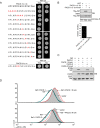
Distinct subsites in the PACS-1 FBR interact with Nef, furin, CK2β, and GGA3. (A) Top, schematic of PACS-1 depicting the atrophin-1–related region (ARR), FBR, middle region (MR) and C-terminal region (CTR), as well as the autoregulation site and sites required for binding AP-1, CK2, and GGA3. Middle, schematic of PACS-1 FBR deletion constructs tested in the two-hybrid screen. Left, amino acid residues; right, interaction of each construct with Nef (+, interaction; –, no interaction). Bottom, sequence alignment of PACS-1181-195 with homologous PACS-2102-116. Nonidentical residues are boxed. (B) Yeast cotransformed with bait plasmids expressing the indicated PACS-1 fragments and prey plasmids expressing Nef, the furin cytosolic domain containing S773S775→DD phosphomimetic mutations (furin-DD), CK2β, or GGA3 were screened for growth on His+ or His media supplemented with 5 mM 3AT. (C) GST-PACS-1181-195, GST-PACS-1117-266, or GST alone was incubated with His6-Nef or His6-fur in-DD. GST proteins were captured, and bound His6-Nef or His6-furin-DD was detected by Western blot. Interactions were assayed in triplicate, and results are presented as the mean ± SD.
FIGURE 2:
FIGURE 2:
PACS-1 N188 and PACS-2 N109 are required for interacting with Nef and for Nef-induced MHC-I down-regulation. (A) Top, yeast cotransformed with bait plasmids expressing PACS-1181-195 containing stepwise alanine mutations (mutated residues colored red) and a prey plasmid expressing full-length Nef (Nef1-206) were screened for growth on His+ or His media supplemented with 5 mM 3AT. Bottom, yeast cotransformed with a bait plasmid expressing PACS-2102-116 and a prey plasmid expressing full-length Nef were screened for growth on His+ or His media supplemented with 5 mM 3AT. (B) GST-PACS-1181-195, GST-PACS-1181-195N188A, or GST alone was incubated with His6-Nef. GST proteins were captured, and bound His6-Nef was detected by Western blot. Protein capture was assayed in triplicate, and results are presented as the mean ± SD. (C) HeLa cells expressing Nef-eYFP alone or coexpressing Nef-eYFP and the indicated HA-tagged PACS proteins were lysed and HA-tagged proteins immunoprecipitated; coimmunoprecipitating Nef was detected by Western blot. (D) Top, H9 cells were nucleofected with plasmids expressing eYFP (vector, gray) or Nef-eYFP alone (Nef, black) or coexpressing Nef-eYFP and PACS-1 (blue) or PACS-1N188A (red). At 40 h postnucleofection, cultures were fixed and eYFP+ cells analyzed for cell-surface MHC-I (mAb W6/32) by flow cytometry. Bottom, H9 cells were nucleofected with plasmids expressing eYFP (vector, gray) or Nef-eYFP alone (Nef, black) or coexpressing Nef-eYFP and PACS-2 (cyan) or PACS-2N109A (red). At 40 h postnucleofection, cultures were fixed and eYFP+ cells analyzed for cell-surface MHC-I (mAb W6/32) by flow cytometry.
FIGURE 3:
FIGURE 3:
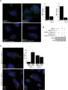
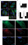
Nef interacts with PACS-1 and PACS-2 on distinct endosome populations. (A) Left, HeLa cells coexpressing Nef-Yc and PACS-1-Yn, PACS-2-Yn, PACS-1N188A-Yn, or PACS-2N109A-Yn for 24 h were fixed and counterstained with DAPI (blue), and the resulting BiFC signal (green) was captured on a high-resolution, wide-field Core DeltaVision system. Scale bar, 10 μm. Right, the BiFC signals generated by coexpression of Nef-Yc with PACS-1-Yn, PACS-2-Yn, PACS-1N188A-Yn, or PACS-2N109A-Yn from a minimum of 100 cells in three independent experiments were determined as described in Materials and Methods and quantified and presented as the mean ± SD. (B) HeLa cells coexpressing the indicated constructs were harvested, and the level of Nef-Yc and PACS-1-Yn, PACS-2-Yn, PACS-1N188A-Yn, or PACS-2N109A-Yn was measured by Western blot. (C) HeLa cells coexpressing Nef-Yc together with PACS-1-Yn and mcherry-Rab7 (upper left) or Nef-Yc together with PACS-2-Yn and either mcherry-Rab7 (lower left) or mcherry-Rab5 (lower right) for 24 h were fixed, counterstained with DAPI (blue), and analyzed for BiFC (green) or Rab proteins (red) using a high-resolution, wide-field Core DeltaVision system. Scale bar, 10 μm. Upper right, quantitation. The colocalization of the BiFC signal generated by coexpression of Nef-Yc with PACS-1-Yn or PACS-2-Yn with mcherry-tagged Rab5 or Rab7 proteins from a minimum of 35 cells in four independent experiments was quantified as described in Materials and Methods and is presented as the mean ± SD.
FIGURE 4:
FIGURE 4:
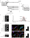
Nef EEEE65 and Pro78 are required for the interaction with PACS-1 and PACS-2. (A) Top, schematic of Nef depicting the M20, EEEE65, PXXP75, and P78 sites required for MHC-I down-regulation. Bottom, schematic of the Nef deletion constructs tested in the two-hybrid screen. Amino acid residues are presented in the stick diagrams, and interaction of each construct with PACS-1181-195 is presented on the right (+, interaction; –, no interaction). (B–D) Yeast cotransformed with the bait plasmid expressing PACS-1181-195 and prey plasmids expressing full-length Nef or the indicated Nef mutants were screened for growth on His+ or His media supplemented with 5 mM 3AT. (E) HeLa cells coexpressing eYFP-tagged Nef or NefE4AP78A with either HA-tagged PACS-1 or PACS-2 as indicated were lysed, HA-tagged proteins immunoprecipitated, and coimmunoprecipitating Nef or NefE4AP78A was detected by western blot. (F) H9 cells were nucleofected with plasmids expressing eYFP (vector, gray), Nef-eYFP (Nef, black), or NefE4AP78A-eYFP (NefE4AP78A, red). At 40 h postnucleofection, cultures were fixed and eYFP+ cells analyzed for cell-surface MHC-I (mAb W6/32) by flow cytometry. (G) Top, HeLa cells expressing Nef-eYFP or NefE4AP78A-eYFP for 40 h were fixed, incubated with anti-TfR (red), and counterstained with DAPI (blue). At 40 h postnucleofection, cells were fixed and stained for TfR (red), and images were captured using a high-resolution, wide-field Core DeltaVision system. Scale bar, 10 μm. Bottom, HeLa cells were transfected with a nonspecific siRNA (NS) or siRNA directed against PACS-2 for 24 h and then nucleofected with a plasmid expressing Nef-eYFP. At 40 h postnucleofection, cells were analyzed as described. Insets, magnification of boxed areas. Right, colocalization of TfR with Nef-eYFP or NefE4AP78A-eYFP was quantified as described in Materials and Methods. Error bars represent the mean ± SD from 30 cells in three independent experiments.
FIGURE 5:
FIGURE 5:
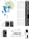
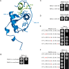
Interaction of the PACS-1 FBR with Nef helix αB. (A) Molecular docking of PACS-1181-195 with Nef (PDB ID: 2Nef, blue). Light green surface images represent an ensemble of the cluster with the highest number of poses for the docking of PACS-1181-195 to Nef. Nef EEEE65 (red) and P78 and Y120 (sticks) are highlighted. (B) Yeast cotransformed with bait plasmids expressing PACS-1181-195 (left) or ARF1T31N (right) and prey plasmids expressing the indicated Nef constructs were screened for growth on His+ or His media supplemented with 50 mM 3AT. (C) Yeast cotransformed with the bait plasmid expressing PACS-1181-195 and prey plasmids expressing the indicated Nef constructs were screened for growth on His+ or His media supplemented with 50 mM 3AT. (D) Yeast cotransformed with a bait plasmid expressing PACS-1181-195 and prey plasmids expressing Nef residues 104–119 containing stepwise alanine mutations (mutated residues colored red) were screened for growth on His+ or His media supplemented with 50 mM 3AT. (E) GST-PACS-1181-195, GST-ARF1 T31N, or GST alone was incubated with His6-Nef or His6-NefE4AW113A. GST proteins were captured, and bound His6-Nef or His6-NefE4AW113A was detected by Western blot. Interactions were assayed in triplicate, and results are presented as the mean ± SD.
FIGURE 6:
FIGURE 6:
PACS-1 interacts with Nef EEEE65 and W113. (A) HeLa cells were nucleofected with plasmids coexpressing HA-tagged PACS-1 or PACS-2 with eYFP-tagged Nef, NefE4A, or NefE4AW113A. After 24 h, cells were lysed and HA-tagged proteins immunoprecipitated; coimmunoprecipitating Nef proteins were detected by Western blot. (B) H9 cells were nucleofected with plasmids expressing eYFP (vector, gray), Nef-eYFP (Nef, black), or NefE4AW113A-eYFP (NefE4AP78A, red). At 40 h postnucleofection, cultures were fixed and eYFP+ cells analyzed for cell-surface MHC-I (mAb W6/32) by flow cytometry.
FIGURE 7:
FIGURE 7:
Mutation of the PACS-binding site disrupts Nef trafficking. (A) Top, HeLa cells expressing Nef-eYFP or NefE4AW113A-eYFP for 40 h were fixed, incubated with anti-TfR (red), and counterstained with DAPI (blue), and images were captured using a high-resolution, wide-field Core DeltaVision system. Middle, HeLa cells were nucleofected with plasmids expressing Nef-eYFP or NefE4AW113A-eYFP alone or coexpressing the eYFP-tagged Nef constructs with either mcherry-Rab5 or mcherry-Rab7 as indicated. At 40 h postnucleofection, cells were fixed, incubated with anti-TfR (red) or not as indicated, and counterstained with DAPI (blue), and images were captured as described. Scale bar, 10 μm. Bottom, magnification of the boxed region from the panel above (right), together with the black and white images of the NefE4AW113A-eYFP signal (middle) and the mcherry-Rab5 signal (right). Blue arrowheads, NefE4AW113A-eYFP. Yellow arrowheads, mcherry-Rab5. (B) Colocalization of Nef-eYFP or NefE4AW113A-eYFP with each endocytic marker was determined as described in Materials and Methods. Error bars represent the mean ± SD from at least 50 cells from three (mcherry-tagged Rab 5 or Rab7) or four (TfR) independent experiments. (C) Colocalization of mcherry-tagged Rab5 or Rab7 with NefE4AW113A-eYFP–positive tubules (left graph) or donut-shaped structures (right graph) was quantified as described in Materials and Methods. Error bars represent the mean ± SD of at least 300 tubule or donut structures from three independent experiments. (D) HeLa cells from experiments described in A coexpressing NefE4AW113A-eYFP (top) and mcherry-Rab5 (middle) or mcherry-Rab7 (middle) for 24 h were fixed and counterstained with DAPI (blue). Images were captured using structured illumination microscopy, and 3D surface representations were generated using Imaris (bottom). Representative Nef-E4AW113A-eYFP–containing tubular (top left) or donut-shaped (top right) structures are highlighted with arrowheads. The arrowheads were then copied on to the middle images showing the mcherry-tagged Rab5 (left) or Rab7 (right) channels to maintain selection of the same structures. Highlighted compartments are pseudocolored for NefE4AW113A-eYFP (cyan) or mcherry-Rab proteins (magenta). Scale bar, 5 μm. The discrete compartments depicted in the model of tubular Nef-E4AW113A-eYFP likely reflect varicosities along the tubule enriched in Nef-E4AW113A-eYFP, since the tubules are frequently continuous in the raw image. The apparent breaks in the tubules reflect the signal detection limits used for the 3D image processing.
FIGURE 8.
FIGURE 8.
Identification of bipartite binding sites in PACS-1 and Nef that mediate protein–protein interaction. (A) Molecular docking of PACS-1181-195 (green) with Nef (PDB ID: 2Nef, blue). PACS-1 amino acids R185 and N188 and Nef residues E64, P78, W113, and Y120 (sticks) are highlighted. (B–D) Yeast cotransformed with plasmids expressing Nef104-119 and PACS-1181-195N188A or PACS-1181-195N188Q (panel B), PACS-1181-195, and Nef104-119W113F or Nef104-119W113A (C) or PACS-1181-195N188Q and Nef104-119W113F (D) were screened for growth on His+ or His media supplemented with 50 mM 3AT. (E, F) Yeast cotransformed with the prey plasmids expressing Nef1-65 (E) or Nef104-119 (F) and bait plasmids expressing the indicated PACS-1 alanine mutants (red) were screened for growth on His+ or His media supplemented with 5 mM 3AT.

References

    1. Ali SA, Huang MB, Campbell PE, Roth WW, Campbell T, Khan M, Newman G, Villinger F, Powell MD, Bond VC. Genetic characterization of HIV type 1 Nef-induced vesicle secretion. AIDS Res Hum Retroviruses. 2010;26:173–192. - PMC - PubMed
    1. Arold S, Franken P, Strub MP, Hoh F, Benichou S, Benarous R, Dumas C. The crystal structure of HIV-1 Nef protein bound to the Fyn kinase SH3 domain suggests a role for this complex in altered T cell receptor signaling. Structure. 1997;5:1361–1372. - PubMed
    1. Aslan JE, et al. Akt and 14-3-3 control a PACS-2 homeostatic switch that integrates membrane traffic with TRAIL-induced apoptosis. Mol Cell. 2009;34:497–509. - PMC - PubMed
    1. Atkins KM, Thomas L, Youker RT, Harriff MJ, Pissani F, You H, Thomas G. HIV-1 Nef binds PACS-2 to assemble a multikinase cascade that triggers major histocompatibility complex class I (MHC-I) down-regulation: analysis using short interfering RNA and knock-out mice. J Biol Chem. 2008;283:11772–11784. - PMC - PubMed
    1. Blagoveshchenskaya AD, Thomas L, Feliciangeli SF, Hung CH, Thomas G. HIV-1 Nef down-regulates MHC-I by a PACS-1- and PI3K-regulated ARF6 endocytic pathway. Cell. 2002;111:853–866. - PubMed

Publication types

MeSH terms

Substances