. 2012 Jun;23(11):2066-75.
doi: 10.1091/mbc.E11-10-0884. Epub 2012 Apr 11.

Lamin B1 loss is a senescence-associated biomarker

Affiliations

Lamin B1 loss is a senescence-associated biomarker

Adam Freund et al. Mol Biol Cell. 2012 Jun.

Abstract

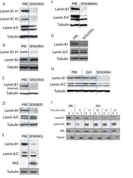
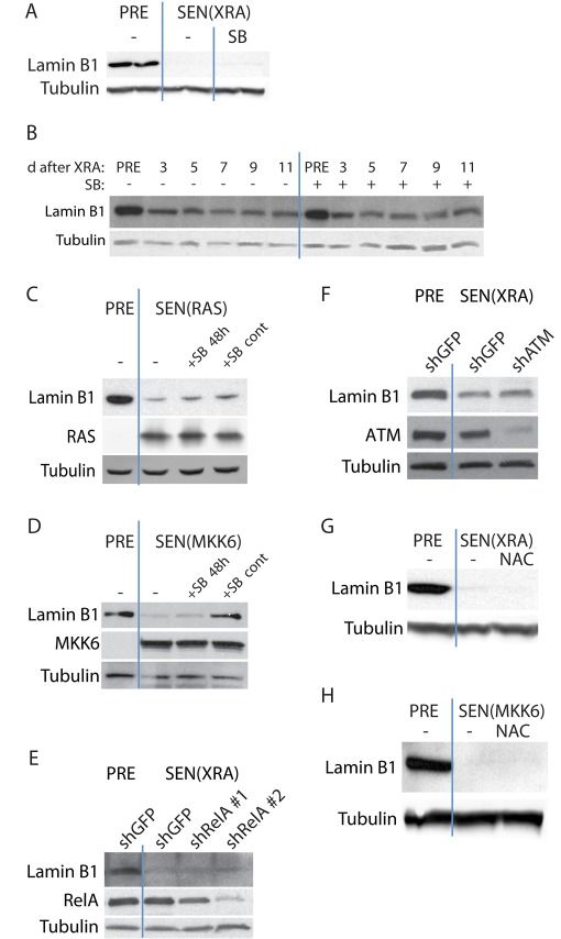
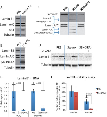
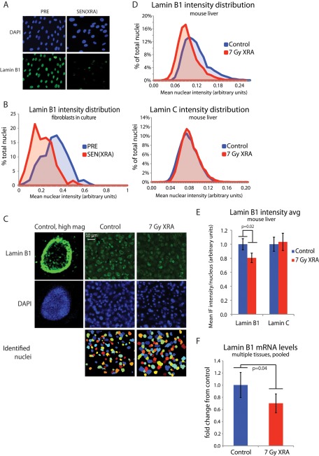
Cellular senescence is a potent tumor-suppressive mechanism that arrests cell proliferation and has been linked to aging. However, studies of senescence have been impeded by the lack of simple, exclusive biomarkers of the senescent state. Senescent cells develop characteristic morphological changes, which include enlarged and often irregular nuclei and chromatin reorganization. Because alterations to the nuclear lamina can affect both nuclear morphology and gene expression, we examined the nuclear lamina of senescent cells. We show here than lamin B1 is lost from primary human and murine cell strains when they are induced to senesce by DNA damage, replicative exhaustion, or oncogene expression. Lamin B1 loss did not depend on the p38 mitogen-activated protein kinase, nuclear factor-κB, ataxia telangiectasia-mutated kinase, or reactive oxygen species signaling pathways, which are positive regulators of senescent phenotypes. However, activation of either the p53 or pRB tumor suppressor pathway was sufficient to induce lamin B1 loss. Lamin B1 declined at the mRNA level via a decrease in mRNA stability rather than by the caspase-mediated degradation seen during apoptosis. Last, lamin B1 protein and mRNA declined in mouse tissue after senescence was induced by irradiation. Our findings suggest that lamin B1 loss can serve as biomarker of senescence both in culture and in vivo.


Figures

FIGURE 1:
FIGURE 1:
Lamin B1 loss is associated with multiple types of cellular senescence. (A) Lamin B1 declines in DNA damage–induced senescence. HCA2 cells were mock irradiated (PRE) or irradiated (10-Gy x-rays) and allowed to senesce (SEN(XRA)). Whole-cell lysates were analyzed by Western blotting using either of two unrelated lamin B1 antibodies that recognize C-terminal epitopes. (B) Lamin B2 does not decline in DNA damage–induced senescence. HCA2 cells were mock irradiated (PRE) or irradiated and allowed to senesce (SEN(XRA)). Whole-cell lysates were analyzed by Western blotting. (C) Lamin B1 declines in SEN(XRA) cells. BJ cells were mock irradiated (PRE) or irradiated and allowed to senesce (SEN(XRA)). Whole-cell lysates were analyzed by Western blotting using a third lamin B1 antibody that recognizes an internal epitope. (D) Lamin B1 declines in replicative senescence. HCA2 cells were cultured until replicative senescence (SEN(REP); ∼70 population doublings). Whole-cell lysates were analyzed by Western blotting. (E) Lamin B1 declines in RAS-induced senescence. HCA2 cells were infected with a lentivirus lacking an insert (PRE) or expressing oncogenic RASV12 and allowed to senesce (SEN(RAS)). Whole-cell lysates were analyzed by Western blotting. (F) Lamin B1 declines in MKK6-induced senescence. HCA2 cells were infected with a lentivirus lacking an insert (PRE) or expressing a constitutively active MAP kinase kinase 6 mutant (MKK6EE) and allowed to senesce (SEN(MKK6). Whole-cell lysates were analyzed by Western blotting. (G) Lamin B1 declines in WI-38 cells after XRA. WI-38 cells were irradiated (SEN(XRA)) and allowed to senesce. PRE cells were mock irradiated. Whole-cell lysates were analyzed by Western blotting. (H) Lamin B1 does not decline in quiescent cells. HCA2 cells were cultured in media containing 10% serum (PRE), serum-free media for 48 h to induce quiescence (QUI), or irradiated and allowed to senesce (SEN(XRA)). Whole-cell lysates were analyzed by Western blotting. (I) Lamin B1 declines within 48 h after induction of senescence by irradiation. HCA2 cells were mock irradiated (PRE) or irradiated (XRA). Nuclear (N) and cytoplasmic (C) extracts collected at the indicated time points were analyzed by Western blotting. RPA serves as a loading control for the nuclear fraction; tubulin serves as a loading control for the cytoplasmic fraction.
FIGURE 2:
FIGURE 2:
Lamin B1 loss at senescence is independent of p38 MAPK, NF-κB, ATM, and ROS signaling. (A) p38 MAPK inhibition does not reverse lamin B1 decline in DNA damage–induced senescence. The p38 MAPK inhibitor SB203580 (SB; 10 μM) was added to SEN(XRA) HCA2 cells for 48 h. Whole-cell lysates were analyzed by Western blotting. PRE cells were mock irradiated. (B) p38 MAPK inhibition does not prevent lamin B1 decline in DNA damage–induced senescence. SB was added to HCA2 cells before XRA. Cells were mock irradiated (PRE) or irradiated (XRA). Whole-cell lysates collected at the indicated time points were analyzed by Western blotting. SB was replaced daily. (C) p38 MAPK inhibition does not reverse or prevent RAS-induced lamin B1 decline. HCA2 cells were infected with a lentivirus expressing oncogenic RASV12 and allowed to senesce (SEN(RAS)). SB was not added (–), added 8 d after infection for 48 h (+SB 48 h), or added before infection and maintained for 10 d (+SB cont). Presenescent controls (PRE) were infected with an insertless vector. Whole-cell lysates were analyzed by Western blotting. (D) p38 MAPK inhibition prevents but does not reverse MKK6-induced lamin B1 decline. HCA2 cells were infected with a lentivirus expressing MKK6EE and allowed to senesce (SEN(MKK6)). SB was not added (–), added 8 d after infection for 48 h (+SB 48 h), or added before infection and maintained for 10 d (+SB cont). Presenescent controls (PRE) were infected with an insertless vector. Whole-cell lysates were analyzed by Western blotting. (E) RelA depletion does not prevent DNA damage–induced lamin B1 decline. HCA2 cells were infected with a lentivirus expressing either of two shRNAs against RelA (shRelA) or GFP (shGFP; control) and selected. Cells were then irradiated and allowed to senesce (SEN(XRA)). Presenescent controls (PRE) were mock irradiated. Whole-cell lysates were analyzed by Western blotting. (F) ATM depletion does not prevent DNA damage–induced lamin B1 decline. HCA2 cells were infected with a lentivirus expressing an shRNA against ATM (shATM) or GFP (shGFP; control) and selected. Cells were then irradiated and allowed to senesce (SEN(XRA)). Presenescent controls (PRE) were mock irradiated. Whole-cell lysates were analyzed by Western blotting. (G) ROS inhibition does not prevent DNA damage–induced lamin B1 decline. NAC (10 mM) was added to HCA2 cells before irradiation and maintained until whole-cell lysates were collected 10 d after XRA (SEN(XRA)). Presenescent controls (PRE) were mock irradiated. NAC was replaced daily. Whole-cell lysates were analyzed by Western blotting. (H) ROS inhibition does not prevent MKK6-induced lamin B1 decline. NAC (10 mM) was added to HCA2 cells before infection and continued until sample collection. Whole-cell lysates were collected 10 d after infection with a lentivirus expressing MKK6EE (SEN(MKK6)). Presenescent controls (PRE) were infected with a lentivirus lacking an insert. NAC was replaced daily. Whole-cell lysates were analyzed by Western blotting.
FIGURE 3:
FIGURE 3:
p53 or p16 expression is sufficient to cause lamin B1 loss, which is regulated at the mRNA level. (A) p53 stabilization is sufficient to cause lamin B1 decline. HCA2 cells were treated with 5 μM nutlin-3a or vehicle (PRE). After 4 d of continuous treatment, whole-cell lysates were analyzed by Western blotting. (B) Ectopic p16INK4A expression is sufficient to cause lamin B1 decline. HCA2 cells were infected with a lentivirus lacking an insert (PRE) or expressing p16INK4A (p16INK4a OE) and selected. Four days after infection, whole-cell lysates were analyzed by Western blotting. (C) Lamin cleavage products are present in apoptotic but not senescent cells. HCA2 cells were treated with 500 nM staurosporine for 24 h to induce apoptosis (Stauro) or irradiated and collected 4 d later (SEN(XRA)). Presenescent controls (PRE) were mock irradiated. Whole-cell lysates were analyzed by Western blotting. (D) Caspase inhibition prevents staurosporine-induced lamin B1 degradation but not senescence-associated lamin B1 decline. HCA2 cells were treated with 500 nM staurosporine for 24 h (Stauro) or irradiated and collected 4 d later (SEN(XRA)). Presenescent controls (PRE) were mock irradiated. Where indicated, the pan-caspase inhibitor z-VAD-fmk (Z-VAD; 100 μM) was added starting before staurosporine/XRA and continuing until whole-cell lysates were collected. Z-VAD-fmk was replaced daily. (E) Lamin B1 mRNA declines in senescent cells. HCA2 and IMR-90 cells were mock irradiated (PRE) or treated with XRA. Total RNA was isolated 4 d later and analyzed by quantitative reverse transcription-PCR. The signal was normalized to tubulin. (F) Lamin B1 mRNA stability is decreased in senescent cells. HCA2 cells were mock irradiated (PRE) or treated with XRA. Three days later, actinomycin D was added (1 μg/ml final) to halt transcription. After 24 h, total RNA was isolated and analyzed by quantitative RT-PCR. Transcript levels immediately before actinomycin D addition were set to 100%.
FIGURE 4:
FIGURE 4:
Lamin B1 loss occurs in vivo after acute DNA damage. (A) Representative images of lamin B1 immunofluorescence. HCA2 cells were mock irradiated (PRE) or irradiated (XRA) and allowed to senesce (SEN(XRA)). Cells were fixed and immunostained for lamin B1. (B) Validation of lamin B1 quantitation via immunofluorescence: histogram of nuclear intensity. HCA2 cells were treated as in A. Lamin B1 intensity was measured across each identified nuclear area using Cell Profiler. p < 0.0001 (C) Representative images of lamin B1 staining in mouse liver. Mice were irradiated (7-Gy XRA) or untreated (control). Twelve weeks later, livers were harvested, sectioned, and immunostained for lamin B1. Lamin B1 intensity was measured across each identified nuclear area. (D) Lamin B1 but not lamin C nuclear intensity declines in mouse liver after irradiation: histogram of nuclear lamin intensity. Mice were irradiated or untreated (control). Twelve weeks later, livers were harvested, sectioned, and immunostained for lamin B1 (top) and lamin C (bottom). Nuclear staining was quantitated with Cell Profiler. For both control and XRA conditions, bin populations were calculated by averaging the relative contribution from each of four mice. (E) Lamin B1 but not lamin C nuclear intensity declines in mouse liver after irradiation: average nuclear lamin intensity. Data from D are presented as average of the mean lamin intensity from four mice for both control and XRA conditions. p = 0.02 for lamin B1, p = 0.71 for lamin C. (F) Lamin B1 mRNA declines in vivo after irradiation. Mouse liver, lung, kidney, and skin were harvested from control mice and mice 12 wk after irradiation. mRNA was purified and lamin B1 mRNA levels determined by quantitative PCR, normalized to tubulin. To generate a single lamin B1 mRNA value from four organs for each mouse, each organ was weighted equally (average control lamin B1 level of each organ set to 1) and pooled for each mouse. Whole-mouse pools were then averaged for each condition: control, n = 4; 7-Gy XRA, n = 5.

Similar articles

Cited by

References

    1. Barascu A, Le Chalony C, Pennarun G, Genet D, Imam N, Lopez B, Bertrand P. Oxidative stress induces an ATM-independent senescence pathway through p38 MAPK-mediated lamin B1 accumulation. EMBO J. 2012;31:1080–1094. - PMC - PubMed
    1. Bargonetti J, Manfredi JJ. Multiple roles of the tumor suppressor p53. Curr Opin Oncol. 2002;14:86–91. - PubMed
    1. Beausejour CM, Krtolica A, Galimi F, Narita M, Lowe SW, Yaswen P, Campisi J. Reversal of human cellular senescence: roles of the p53 and p16 pathways. EMBO J. 2003;22:4212–4222. - PMC - PubMed
    1. Benson EK, Lee SW, Aaronson SA. Role of progerin-induced telomere dysfunction in HGPS premature cellular senescence. J Cell Sci. 2010;123:2605–2612. - PMC - PubMed
    1. Bridger JM, Foeger N, Kill IR, Herrmann H. The nuclear lamina. Both a structural framework and a platform for genome organization. FEBS J. 2007;274:1354–1361. - PubMed

Publication types

MeSH terms