. 2012 Jun;23(11):2028-40.
doi: 10.1091/mbc.E11-11-0911. Epub 2012 Apr 11.

NHERF1 acts as a molecular switch to program metastatic behavior and organotropism via its PDZ domains

Affiliations

NHERF1 acts as a molecular switch to program metastatic behavior and organotropism via its PDZ domains

Rosa Angela Cardone et al. Mol Biol Cell. 2012 Jun.

Abstract

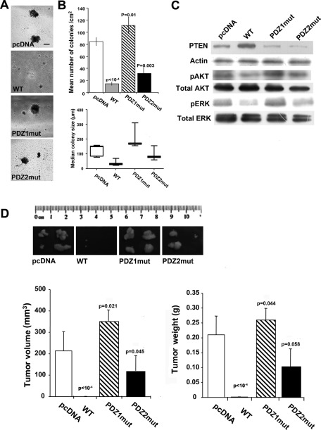
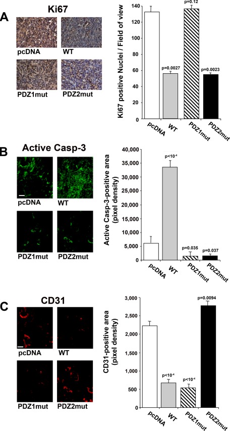
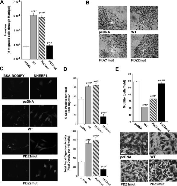
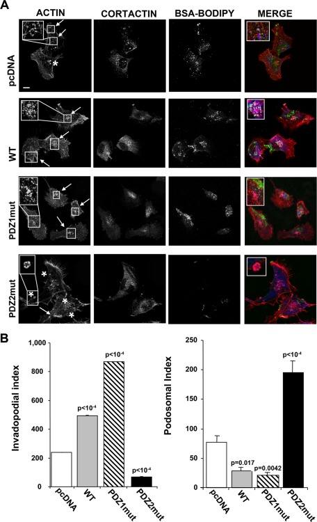
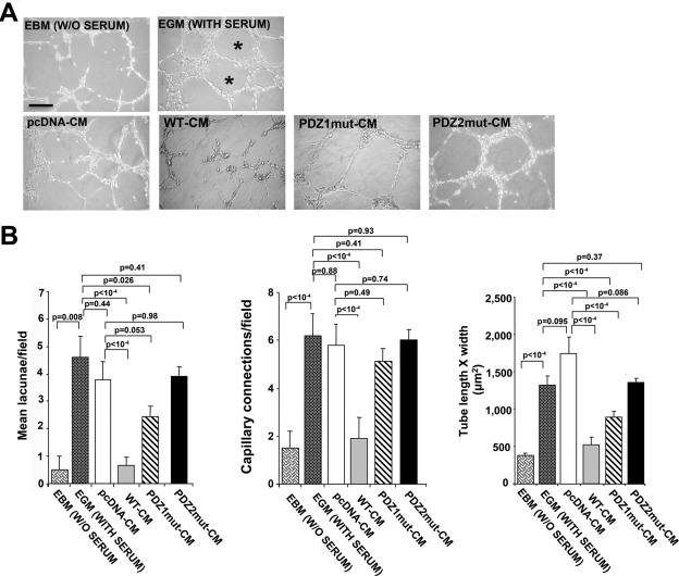
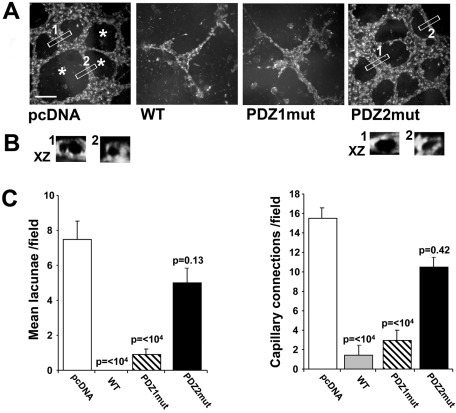
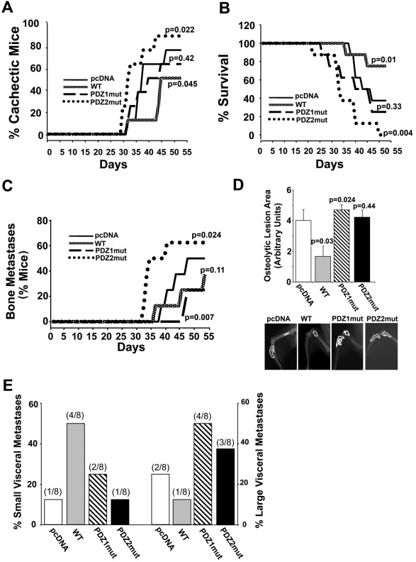
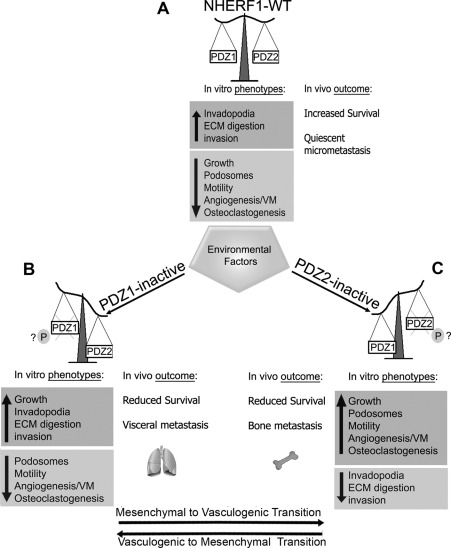
Metastatic cells are highly plastic for differential expression of tumor phenotype hallmarks and metastatic organotropism. The signaling proteins orchestrating the shift of one cell phenotype and organ pattern to another are little known. Na(+)/H(+) exchanger regulatory factor (NHERF1) is a molecular pathway organizer, PDZ-domain protein that recruits membrane, cytoplasmic, and cytoskeletal signaling proteins into functional complexes. To gain insight into the role of NHERF1 in metastatic progression, we stably transfected a metastatic breast cell line, MDA-MB-231, with an empty vector, with wild-type NHERF1, or with NHERF1 mutated in either the PDZ1- or PDZ2-binding domains to block their binding activities. We observed that NHERF1 differentially regulates the expression of two phenotypic programs through its PDZ domains, and these programs form the mechanistic basis for metastatic organotropism. The PDZ2 domain promotes visceral metastases via increased invadopodia-dependent invasion and anchorage-independent growth, as well as by inhibition of apoptosis, whereas the PDZ1 domain promotes bone metastases by stimulating podosome nucleation, motility, neoangiogenesis, vasculogenic mimicry, and osteoclastogenesis in the absence of increased growth or invasion. Collectively, these findings identify NHERF1 as an important signaling nexus for coordinating cell structure with metastatic behavior and identifies the "mesenchymal-to-vasculogenic" phenotypic transition as an essential step in metastatic progression.


Figures

FIGURE 1:
FIGURE 1:
Increased expression of NHERF1 impairs tumor growth mainly via the PDZ1 domain. (A–C) MDA-MB-231 cells transfected with empty vector (pcDNA), WT NHERF1 (WT), and the PDZ1 (PDZ1mut) and PDZ2 (PDZ2mut) mutated constructs were plated onto soft agar. After 4 wk, colonies were counted, and colony size was assessed by micrometer. (A) Representative photomicrographs of colonies for each cell variant. Results are represented as mean ± SEM; the values of p are vs. pcDNA cells. (B) Top, number of colonies measuring >50 μm; bottom, median size of colonies, with the bars representing the minimum and maximum values for each clone. (C) Western blot for the levels of PTEN and of phospho-ERK and phospho-AKT kinases using the total and phospho-specific antibodies listed in Materials and Methods. (D) In vivo tumor growth. Female BALB/c-nu/nu mice were subcutaneously injected with cell variants as indicated. Top, photographs of four tumors per cell variant at the day of dissection. Bottom, tumor volume and weight measured at 32 d postinjection; n = 4 mice/group; results are represented as mean ± SEM; the values of p are vs. pcDNA-injected animals.
FIGURE 2.
FIGURE 2.
NHERF1 regulates tumor cell proliferation and apoptosis. Tumor sections from tumors obtained by subcutaneous injection of pcDNA-, WT-, PDZ1mut-, and PDZ2mut-transfected cells were subjected to (A) immunohistochemical analysis for the proliferative marker Ki-67, (B) immunofluorescence assay for the apoptosis marker active caspase-3, and (C) the endothelial marker CD31. Left, representative images at the original magnification (bar, 10 μm). Right, quantification of expression performed using ImageJ software, n = 4; results are represented as mean ± SEM; the values of p are compared with levels in pcDNA tumors.
FIGURE 3:
FIGURE 3:
The lack of functional PDZ2 domain decreases invasion of tumor cells. (A) The ability of the indicated transfected MDA-MB-231 cells to cross a Matrigel layer in 6 h was analyzed in Boyden chambers. Each cell variant was evaluated five times in triplicate, and results are represented as mean ± SEM. The values of p compared with the pcDNA-transfected cells. (B) Representative microphotographs of Matrigel evasion assays. Cells were included in growth factor–free Matrigel drops and incubated at 37°C for 6 d (bar, 30 μm). (C) NHERF1 regulation of ECM degradation measured by an in vitro fluorescence–Matrigel degradation assay. Cells were seeded on coverslips coated with Matrigel (4 mg/ml) and BSA-BODIPY (30 μg/ml) and incubated at 37°C for 24 h. Panels show representative pictures of proteolytic degradation in the Matrigel matrix in green (BSA-BODIPY) and immunofluorescence of NHERF1 protein expression in red (NHERF1; bar, 10 μm). (D) Quantification of invadopodia function (ECM degradation) analyzed as percentage of cells digesting the matrix (top) or total focal digestive activity of 100 cells (bottom); n = 5; mean ± SEM values of p test vs. pcDNA. (E) Migration assay of the indicated cell lines through collagen IV–coated filters in Boyden chambers. Results are expressed as numbers of migrated cells/field. Top, representative fields of migrated cells through filters. Bar, 10 μm. Mean ± SEM of three independent experiments performed in duplicate. The values of p are compared with the pcDNA-transfected cells.
FIGURE 4:
FIGURE 4:
The PDZ2 domain regulates invadopodia and the PDZ1 domain regulates podosome dynamics in NHERF1-overexpressing cells. Invadopodia and podosome rings in the cells cultured on glass coverslips coated with Matrigel containing BSA-BODIPY for 24 h to visualize ECM digestion (green) and stained for F-actin (red) and cortactin (blue). (A) Confocal immunofluorescence localization shows invadopodia (arrows) visible as F-actin dots colocalizing with cortactin and overlapping with areas of ECM degradation. Podosomal ring structures are marked with cortactin and surround F-actin rich cores in pcDNA and PDZ2mut cells (asterisk). Bar, 10 μm. (B) Quantification of the Invadopodia Index (left) and Podosomal Index (right) for each cell variant calculated as described in Materials and Methods. Data are representative or the mean ± SEM of cells from three independent experiments; the values of p are compared with the pcDNA-transfected cells.
FIGURE 5:
FIGURE 5:
NHERF1 inhibits neoangiogenesis via the PDZ2 domain. (A) Representative microphotographs of capillary network formation of HUVECs seeded on Matrigel and incubated with EBM (medium without serum), EGM (medium with serum), or CM of each cell variant for 24 h. Asterisks indicate empty areas (lacunae) bordered by the capillary-like network. (B) Graphs showing quantification of mean number of lacunae/field (left) and of capillary-like connections/field (center), and tube morphology (tube length × width; right) in HUVECs in the presence of EBM, EGM, or CM from each cell variant. Mean ± SEM; the values of p compared with the EBM-, EGM-, or pcDNA-transfected cells as indicated.
FIGURE 6:
FIGURE 6:
NHERF1 inhibits vasculogenic mimicry–like ability via the PDZ2 domain. (A) Representative microphotographs of capillary-like tubule formations obtained with the cells when seeded inside Matrigel for 5 d in their growth medium. Cells were stained with Oregon Green 488 DHPE and acquired by confocal microscopy. Asterisks indicate empty areas (lacunae) bordered by the capillary-like network. Bar, 10 μm. (B) Bottom, XZ-zoomed vertical cross-section views of 3D reconstruction of Z-stacked VM tubes of the regions of interest, indicated by 1 or 2, in each field from pcDNA cells and PDZ2mut cells. Vertical sections of the 3D-reconstructed tubes show that they are open lumen-like structures surrounded by fluorescent cells. (C) The mean number of lacunae/field (left) and the capillary connections number (right) were quantified. Results are expressed as mean ± SEM; p value vs. pcDNA. All assays were performed in quadruplicate.
FIGURE 7:
FIGURE 7:
NHERF1 PDZ domain function dictates organotropism. Four-week-old female BALB/c-nu/nu mice were inoculated in the left ventricle with a suspension of each cell variant. n = 8 mice/group. Mice were monitored daily for (A) cachexia (decrease of body weight) and (B) survival (p values vs. pcDNA-injected mice are presented) and (C) monitored weekly by x-ray analysis to determine onset and incidence of bone metastasis. p value vs. pcDNA-injected mice. (D) Evaluation of osteolytic area by densitometric analysis of hindlimb x-ray. Bottom, representative radiographic images of one hindlimb that developed an osteolytic lesion for each group. Data are presented as mean ± SEM; p value vs. pcDNA. (E) At the end of the experiment animals were killed and subjected to anatomical dissection to evaluate the incidence of visceral metastases, which were scored, according to the size, as percentage of small (left x-axis) or large (right x-axis) metastases.
FIGURE 8:
FIGURE 8:
Proposed model for NHERF1 PDZ domain-orchestrated switches regulating in vitro tumor phenotypes and in vivo metastatic organotropism. On the basis of its expression, NHERF1 may behave as (A) an oncosuppressor (NHERF1-WT) or (B, C) a metastasis-organotropic–specific protein (NHERF1 PDZ1 or PDZ2inactive). Although it is not yet clear which cellular signal mechanisms are responsible for this functional switch, exposure to an aberrant tumor microenvironment (hypoxic stress, nutrient stress, low extracellular pHe, ECM components) may play a role in affecting NHERF1 posttranslational modifications and/or shuttling from one cellular compartment to another, probably via altered phosphorylation and activity of one of its PDZ domains, thus attenuating its oncosuppressor role and activating specific oncogenic pathways. Moreover, the specific loss of function of one of the two PDZ domains, through the selection of a wide and flexible spectrum of possible hallmark behaviors, regulates the shift from one set of metastatic phenotypes to another and the spread to one specific organ to another (metastatic switch). (B) Specifically, when NHERF1 is overexpressed and PDZ1 is blocked (PDZ1mut cells) a growth program is activated, and visceral organs are preferred for spreading because invadopodia-dependent invasion and growth are up-regulated, whereas podosome/motility/angiogenic/osteoclastogenic programs are inhibited. (C) On the other hand, when PDZ2 is blocked (PDZ2mut cells) bone metastases are promoted because podosome/motility, neoangiogenesis, vasculogenic mimicry–like ability, and osteoclastogenesis are stimulated, whereas the invasive program is turned off. The shifting from one program to the other could be controlled by the well-documented phosphorylation-dependent alteration in PDZ domain function of NHERF1. If the switch between these phenotypic programs is bidirectional due to reversible phosphorylation-dependent alterations of the activity of one or the other PDZ domains, then this could define the phenotypic transition termed the mesenchymal–vasculogenic transition.

References

    1. Accardi R, et al. E6 and E7 from human papillomavirus type 16 cooperate to target the PDZ protein Na/H exchange regulatory factor 1. J Virol. 2011;85:8208–8216. - PMC - PubMed
    1. Arguello F, Baggs RB, Frantz CN. A murine model of experimental metastasis to bone and bone marrow. Cancer Res. 1988;48:6876–6881. - PubMed
    1. Artym VV, Matsumoto K, Mueller SC, Yamada KM. Dynamic membrane remodeling at invadopodia differentiates invadopodia from podosomes. Eur J Cell Biol. 2011;90:172–180. - PMC - PubMed
    1. Basbaum CB, Werb Z. Focalized proteolysis: spatial and temporal regulation of extracellular matrix degradation at the cell surface. Curr Opin Cell Biol. 1996;8:731–738. - PubMed
    1. Bellizzi A, Malfettone A, Cardone RA, Mangia A. NHERF1/EBP50 in breast cancer: clinical perspectives. Breast Care (Basel) 2010;5:86–90. - PMC - PubMed

Publication types

MeSH terms

Substances

LinkOut - more resources