. 2012 Jun;23(11):2156-69.
doi: 10.1091/mbc.E11-10-0844. Epub 2012 Apr 11.

Autophagy modulates dynamics of connexins at the plasma membrane in a ubiquitin-dependent manner

Affiliations

Autophagy modulates dynamics of connexins at the plasma membrane in a ubiquitin-dependent manner

Eloy Bejarano et al. Mol Biol Cell. 2012 Jun.

Abstract

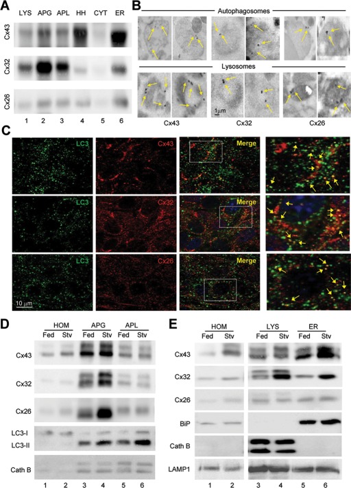
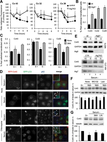
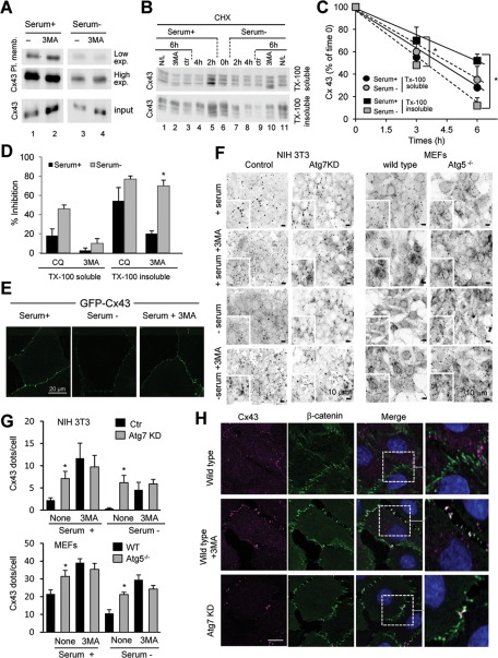
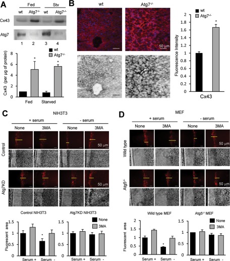
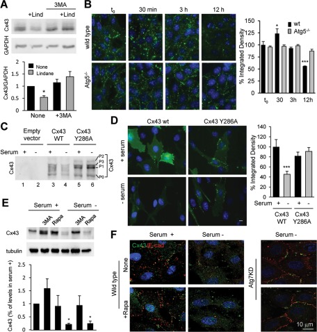
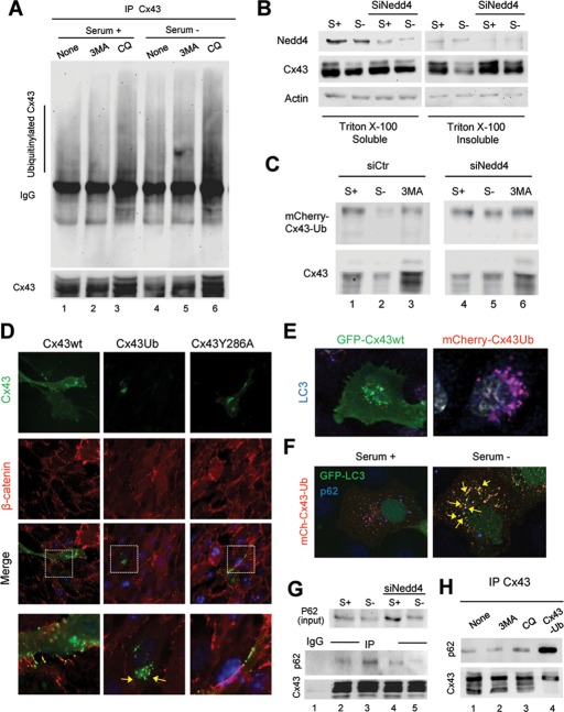
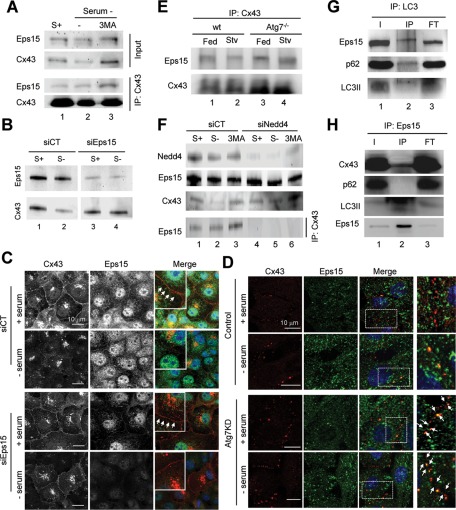
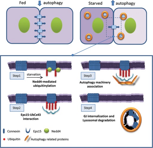
Different pathways contribute to the turnover of connexins, the main structural components of gap junctions (GJs). The cellular pool of connexins targeted to each pathway and the functional consequences of degradation through these degradative pathways are unknown. In this work, we focused on the contribution of macroautophagy to connexin degradation. Using pharmacological and genetic blockage of macroautophagy both in vitro and in vivo, we found that the cellular pool targeted by this autophagic system is primarily the one organized into GJs. Interruption of connexins' macroautophagy resulted in their retention at the plasma membrane in the form of functional GJs and subsequent increased GJ-mediated intercellular diffusion. Up-regulation of macroautophagy alone is not sufficient to induce connexin internalization and degradation. To better understand what factors determine the autophagic degradation of GJ connexins, we analyzed the changes undergone by the fraction of plasma membrane connexin 43 targeted for macroautophagy and the sequence of events that trigger this process. We found that Nedd4-mediated ubiquitinylation of the connexin molecule is required to recruit the adaptor protein Eps15 to the GJ and to initiate the autophagy-dependent internalization and degradation of connexin 43. This study reveals a novel regulatory role for macroautophagy in GJ function that is directly dependent on the ubiquitinylation of plasma membrane connexins.


Figures

FIGURE 1:
FIGURE 1:
Association of connexins with compartments of the autophagic/lysosomal system is modulated by starvation. (A) Immunoblot for the indicated Cx of subcellular fractions (100 μg of protein/lane) isolated from 6-h-starved mice liver. APG, autophagosomes; APL, autophagolysosomes; CYT, cytosol; ER, endoplasmic reticulum; HH, heart homogenate run as positive control for Cx43; LYS, lysosomes; Homogenates are shown in E. (B) Immunogold for the indicated Cxs in lysosomes and autophagosomes isolated from fed mouse liver (complete field images are shown in Supplemental Figure S1C). (C) Immunofluorescence for LC3 and the indicated Cxs in MEF cells. Individual channels, merge, and colocalization regions are shown. Right, a higher-magnification inset; arrows indicate colocalization. (D, E) Immunoblots for the indicated Cxs of autophagic vacuoles (D; APG, autophagosomes; APL, autophagolysosomes) and lysosomes (E; 50 μg of protein/lane) isolated from livers of fed or 6-h-starved (Stv) mice. Controls for purity in each fraction include ER marker (BiP), lysosomal hydrolase (cathepsin B), autophagosomal marker (LC3), and LAMP1.
FIGURE 2:
FIGURE 2:
Degradation of connexins is compromised by blockage of macroautophagy. (A) Kinetics of fluorescence decay in wild-type and Atg7-knockdown NIH3T3 cells transfected with a vector expressing Dendra fused to the indicated Cx imaged at different times after photoactivation. The ratio of red to green fluorescence was calculated at each time point. Values are expressed as percentage of the ratio at time 0 and are mean ± SEM (n = 3). (B, C) MEFs from wild-type and Atg5-null mice (Atg5−/−; B) or NIH3T3 fibroblasts (C) transfected with the same constructs as in A were incubated after photoactivation in the presence or absence of serum and 3MA as labeled. Quantification of red fluorescence decay by FACS analysis calculated as the percentage of total red fluorescence in each sample normalized against the total number of cells expressing Dendra-Cx. Values are mean ± SEM (n = 3). Significant differences with wild-type cells are indicated. (See representative scatterdot plot for each different condition in Supplemental Figure S2.) (D) COS-7 cells transfected with RFP-Cx43 and GFP-LC3 were maintained in the presence or absence of serum and treated or not with chloroquine as labeled. Immunofluorescence for p62 (blue) and the fluorescence in the red (Cx43) and green (LC3) channels is shown. Right, a merged image; inset shows detail at higher magnification. (E) Immunoblot for Cx43 and the indicated Atg in MEFs from wild-type or Atg5-null mice or in NIH3T3 fibroblasts control or knocked down for Atg7 maintained in the presence or absence of serum for 8 h. (F, G) Immunoblots for Cx43 of NIH3T3 cells incubated in the presence (S+) or absence of serum and treated for 8 h with two different proteasome inhibitors (MG132 or lactacystin [Lact]), 3MA, and a combination of leupeptin and ammonium chloride (L/N). Top, representative immunoblots. Bottom, changes in Cx43 levels calculated by densitometric quantification of blots like the ones shown here. Values are expressed as percentage of Cx43 present in cells maintained in serum-supplemented media and are mean ± SEM (n = 3). *p < 0.05. Significant differences with respect to control, wild type, or serum+.
FIGURE 3:
FIGURE 3:
Macroautophagy degrades preferentially Cx43 in GJs at the plasma membrane. (A) COS-7 cells transfected with Cx43 were maintained in the presence or absence of serum and treated or not with 10 mM 3MA as labeled. Cells were then subjected to cell surface protein biotinylation, and the biotinylated fraction of the cell lysates was precipitated with NeutrAvidin beads. Precipitates were then analyzed by Western blot using polyclonal antibodies against Cx43. (B) COS-7 cells transfected with Cx43 were treated with cycloheximide (CHX) for the indicated times either in the presence or absence of serum. Lysates were subjected to extraction with 1% Triton X-100, and soluble and insoluble fractions were processed for immunoblot against Cx43. Where indicated 10 mM 3MA or 20 mM ammonium chloride and 100 μM leupeptin (N/L) was added to the incubation media during the chase. (C) The kinetics of degradation of Cx43 in each condition was calculated by densitometry of immunoblots like the one shown and plotted in a graph. (D) Inhibitory effect of CQ and 3MA in the degradation of Cx43 in each fraction calculated by densitometric quantification of blots like the ones in A. Values are expressed as percentage of inhibition and are mean ± SEM (n = 3). (E) Confocal microscopy of cells expressing GFP-Cx43 maintained in the presence or absence of serum and supplemented with 3MA as indicated. (F) Immunofluorescence (inverted grayscale images) of Cx43 in NIH3T3 cells control or knocked down for Atg7 (left) and in MEFs from wild-type or Atg5-null mice (right) maintained for 8 h in serum-supplemented or serum-deprived media in presence or not of 3MA as indicated. (G) Quantification of number of Cx43-positive fluorescent puncta per cell. Values are mean ± SEM (n = 3; >45 cells counted per experiment). (H) Immunofluorescence for Cx43 and β-catenin in NRK cells control, knocked down for Atg7, or exposed to 10 mM 3MA for 8 h. Individual channels and merged channel images are shown. Right, higher-magnification of insets. *p < 0.05. Significant differences with respect to control or serum+.
FIGURE 4:
FIGURE 4:
Blockage of macroautophagy increases the content of functional GJs. (A) Immunoblot for the indicated proteins of homogenates (50 μg of protein/lane) from livers of fed or 6-h-starved wild-type and Alb-Cre-ATG7f/f mice. Bottom, quantification of enrichment of Cx43 calculated by densitometry of blots like the ones shown here. Values are mean ± SEM (n = 3–5). Significant differences with wild type are indicated. (B) Immunostaining for Cx43 of frozen liver sections from the same mice. Bottom, inverted grayscale images to better visualize GJs. Right, Quantification of changes in fluorescence intensity for Cx43 in the liver sections expressed as fold the intensity in wild type. Values are mean ± SEM (n = 3). (C, D) Scrape loading assay with sulforhodamine B in NIH3T3 cells control or knocked down for Atg7 (C) and MEFs from wild-type mice or mice null for Atg5 (D) incubated in the presence or absence of serum and 10 mM 3MA for 8 h. Histograms at the bottom show the fluorescent area in each condition quantified using Scion Image software relative to the area in control conditions. Values are mean ± SEM (n = 3, with 45 cells counted). *p < 0.05. Significant differences with respect to control.
FIGURE 5:
FIGURE 5:
Blockage of macroautophagy prevents internalization and degradation of Cx43. (A) Immunoblot (100 μg of protein/lane) for Cx43 in MEFs treated with 100 μM lindane (Lind) for 12 h in presence or absence of 10 mM 3MA. Top, representative immunoblot. Bottom, quantification of total levels of Cx43 relative to values in untreated cells. Values are mean ± SEM (n = 5). Significant differences with respect to untreated cells are indicated. (B) Immunofluorescence for Cx43 in MEFs from wild-type and Atg5-null mice at the indicated times after treatment with lindane. Right, quantification of the decay of signal for Cx43 in both cell types. Values are mean ± SEM (n = 3). Significant differences with respect to untreated cells are indicated. (C) Immunoblot for Cx43 of COS-7 cells transfected with Cx43 wild type (wt) or mutated at tyrosine 286 (Cx43Y286A) and maintained in the presence or absence of serum as indicated. (D) Immunofluorescence for Cx43 of COS-7 transfected with plasmids coding for wt and endocytic mutant (Y286A) Cx43 and maintained in the presence or absence of serum as labeled. Left, representative images. Right, quantification of the integrated density of the signal for Cx43 in both cell types. Values are mean ± SEM (n = 3). Significant differences with respect to cells maintained in serum supplemented media are indicated. (E) NIH3T3 cells incubated in the presence or absence of serum and treated for 8 h with 3MA or rapamycin (Rapa). Top, representative immunoblot. Bottom, changes in Cx43 levels calculated by densitometric quantification of blots like the one shown here. Values are expressed as percentage of Cx43 present in untreated cells maintained in serum-supplemented media and are mean ± SEM (n = 3). Significant differences with respect to untreated serum+ are indicated. (F) Immunofluorescence for Cx43 and E-cadherin in cells maintained in the presence or absence of serum and treated or not with rapamycin (Rapa). Merged channels are shown. Right, similar staining in cells knocked down for Atg7. *p < 0.05. Significant differences with respect to control.
FIGURE 6:
FIGURE 6:
Macroautophagy of Cx43 in GJs requires its prior ubiquitinylation. (A) COS-7 cells expressing Cx43 untreated (none) or treated with 10 mM 3-methyladenine or 10 mM chloroquine and maintained in the presence or absence of serum were subjected to immunoprecipitation for Cx43 and immunoblotted for Cx43 or ubiquitin (exposure time on the right has been selected to equal the intensity of the Cx43 bands and enable better appreciation of the differences in intensity of ubiquitinylation). (B) COS-7 cells transfected with Cx43 alone or together with siRNA for Nedd4 were maintained in the presence or absence of serum. Lysates were subjected to extraction with 1% Triton X-100, and soluble and insoluble fractions were processed for immunoblot against the indicated proteins. (C) COS-7 cells transfected with wild-type Cx43 (Cx43WT) or the mCherry-Cx43-Ub plasmid alone or together with siRNA for Nedd4 were maintained in the presence or absence of serum and treated with 3MA where indicated and subjected to immunoblot for Cx43. (D) Immunofluorescence for Cx43 and β-catenin in NRK cells transfected with plasmids coding for wild-type (wt), ubiquitin tagged (-Ub), and endocytic mutant (Y286A) Cx43. Individual channels and merged images are shown. Bottom, insets at higher magnification. (E) Immunostaining for LC3 in NRK cells expressing wild-type GFP-Cx43 or ubiquitin-tagged mCherry-Cx43 maintained in the presence of serum. Merged channels are shown. (F) Immunostaining for p62 of COS-7 cells transiently expressing mCherry-Cx43-Ub and GFP-LC3 and maintained in the presence or absence of serum. Merged channels are shown. (G) Lysates of COS-7 cells transfected with Cx43 and maintained in the presence or absence of serum were subjected to extraction with 1% Triton X-100, and the soluble fraction was subjected to immunoprecipitation for Cx43 and immunoblotted for p62 and Cx43. Levels of p62 in the input are also shown. (H) Lysates from COS-7 cells transfected with Cx43 and/or mCherry-Cx43-Ub, maintained in the absence of serum and treated or not with 10 mM 3MA or 50 μM chloroquine (CQ), were immunoprecipitated with polyclonal antibodies directed against Cx43 and the precipitates immunoblotted for p62 or Cx43.
FIGURE 7:
FIGURE 7:
Degradation of GJ channels via macroautophagy requires binding of Eps15 to Cx. (A) COS-7 cells maintained in the presence (+) or absence (–) of serum and treated with 10 mM 3MA were subjected to immunoprecipitation for Cx43, followed by immunoblot for Cx43 and Eps15. Levels of Eps15 and Cx43 in the input fractions are also shown. (B) Immunoblot for Cx43 and Eps15 of COS-7 cells transfected with a control (CT) siRNA or siRNA for Eps15 and maintained in the presence or absence of serum as labeled. (C) Immunofluorescence for Cx43 (red) and Eps15 (green) in the same cells maintained in the presence or absence of serum as labeled. Single and merged channels are shown. Arrows point to the presence of Cx in the plasma membrane. (D) Immunofluorescence for Cx43 and Eps15 in NRK cells control or knockdown for Atg7 cells incubated in presence or absence of serum for 4 h. Right, higher-magnification images. (E) Immunoblot for Eps15 and Cx43 of immunoprecipitates for Cx43 in livers of wt or Alb-Cre-ATG7f/f (Atg7−/−) mice. (F) Immunoblot for the indicated proteins in cells transfected with a control (CT) siRNA or siRNA for Nedd4 maintained in the presence or absence of serum and treated with 3MA as labeled. Bottom, levels of Eps15 in immuno­precipitates for Cx43 in the same cells. (G) Immunoblot for the indicated proteins in immunoprecipitates for LC3 in starved rat liver. (H) Immunoblot for the indicated proteins in immunoprecipitates for Eps15 in serum-deprived cells. Inputs (I), immunoprecipitates (IP), and flowthrough fraction (FT).
FIGURE 8:
FIGURE 8:
Degradation of GJ channels via macroautophagy requires Nedd4 mediated ubiquitinylation. GJ channels contribute to the metabolic and electrical coupling among adjacent cells. Starvation induces degradation of GJ connexins by macroautophagy and reduces intercellular communication. Macroautophagy of connexins is triggered by their ubiquitinylation by the ubiquitin ligase Nedd4 (step 1). Eps15 associates with the highly enriched ubiquitinylated GJ domains (step 2) and favors recruitment of the autophagic machinery (step 3), leading to connexin internalization and lysosomal degradation (step 4).

References

    1. Berthoud VM, Minogue PJ, Guo J, Williamson EK, Xu X, Ebihara L, Beyer EC. Loss of function and impaired degradation of a cataract-associated mutant connexin50. Eur J Cell Biol. 2003;82:209–221. - PMC - PubMed
    1. Berthoud VM, Minogue PJ, Laing JG, Beyer EC. Pathways for degradation of connexins and gap junctions. Cardiovasc Res. 2004;62:256–267. - PubMed
    1. Bugnicourt A, Mari M, Reggiori F, Haguenauer-Tsapis R, Galan JM. Irs4p and Tax4p: two redundant EH domain proteins involved in autophagy. Traffic. 2008;9:755–769. - PubMed
    1. Catarino S, Ramalho JS, Marques C, Pereira P, Girao H. Ubiquitin-mediated internalization of connexin43 is independent of the canonical endocytic tyrosine-sorting signal. Biochem J. 2011;437:255–267. - PubMed
    1. Csikos G, Lippai M, Lukacsovich T, Juhasz G, Henn L, Erdelyi M, Maroy P, Sass M. A novel role for the Drosophila epsin (lqf): involvement in autophagy. Autophagy. 2009;5:636–648. - PubMed

Publication types

MeSH terms