

Axonal thinning and extensive remyelination without chronic demyelination in spinal injured rats
- PMID: 22496557
- PMCID: PMC3335198
- DOI: 10.1523/JNEUROSCI.0002-12.2012
Axonal thinning and extensive remyelination without chronic demyelination in spinal injured rats
Abstract
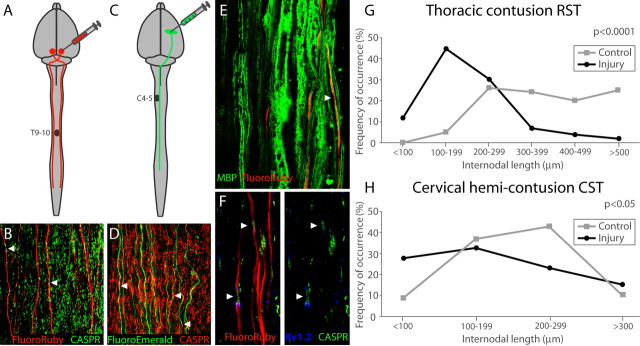
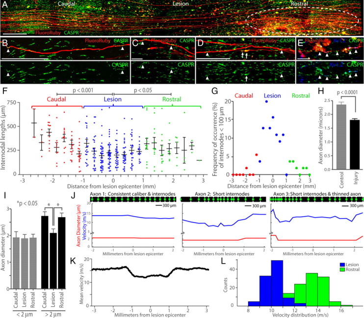
Remyelination following spinal cord injury (SCI) is thought to be incomplete; demyelination is reported to persist chronically and is proposed as a compelling therapeutic target. Yet most reports do not distinguish between the myelin status of intact axons and injury-severed axons whose proximal stumps persist but provide no meaningful function. We previously found full remyelination of spared, intact rubrospinal axons caudal to the lesion in chronic mouse SCI. However, the clinical concept of chronically demyelinated spared axons remains controversial. Since mouse models may have limitations in clinical translation, we asked whether the capacity for full remyelination is conserved in clinically relevant chronic rat SCI. We determined myelin status by examining paranodal protein distribution on anterogradely labeled, intact corticospinal and rubrospinal axons throughout the extent of the lesion. Demyelination was evident on proximal stumps of severed axons, but not on intact axons. For the first time, we demonstrate that a majority of intact axons exhibit remyelination (at least one abnormally short internode, <100 μm). Remarkably, shortened internodes were significantly concentrated at the lesion epicenter and individual axons were thinned by 23% compared with their rostral and caudal zones. Mathematical modeling predicted a 25% decrease in conduction velocity at the lesion epicenter due to short internodes and axonal thinning. In conclusion, we do not find a large chronically demyelinated population to target with remyelination therapies. Interventions may be better focused on correcting structural or molecular abnormalities of regenerated myelin.
Figures
Similar articles
-
No evidence for chronic demyelination in spared axons after spinal cord injury in a mouse.J Neurosci. 2008 Apr 9;28(15):3887-96. doi: 10.1523/JNEUROSCI.4756-07.2008. J Neurosci. 2008. PMID: 18400887 Free PMC article.
-
Cortical stimulation leads to shortened myelin sheaths and increased axonal branching in spared axons after cervical spinal cord injury.Glia. 2023 Aug;71(8):1947-1959. doi: 10.1002/glia.24376. Epub 2023 Apr 25. Glia. 2023. PMID: 37096399 Free PMC article.
-
Compensatory changes after spinal cord injury in a remyelination deficient mouse model.J Neurochem. 2025 Jan;169(1):e16220. doi: 10.1111/jnc.16220. Epub 2024 Sep 13. J Neurochem. 2025. PMID: 39268880 Free PMC article.
-
Remyelination of the injured spinal cord.Prog Brain Res. 2007;161:419-33. doi: 10.1016/S0079-6123(06)61030-3. Prog Brain Res. 2007. PMID: 17618995 Free PMC article. Review.
-
Myelin status and oligodendrocyte lineage cells over time after spinal cord injury: What do we know and what still needs to be unwrapped?Glia. 2019 Nov;67(11):2178-2202. doi: 10.1002/glia.23702. Epub 2019 Aug 24. Glia. 2019. PMID: 31444938 Free PMC article. Review.
Cited by
-
Pathological changes in the white matter after spinal contusion injury in the rat.PLoS One. 2012;7(8):e43484. doi: 10.1371/journal.pone.0043484. Epub 2012 Aug 29. PLoS One. 2012. PMID: 22952690 Free PMC article.
-
Microenvironmental regulation of oligodendrocyte replacement and remyelination in spinal cord injury.J Physiol. 2016 Jul 1;594(13):3539-52. doi: 10.1113/JP270895. Epub 2016 Mar 29. J Physiol. 2016. PMID: 26857216 Free PMC article. Review.
-
Surgical Considerations to Improve Recovery in Acute Spinal Cord Injury.Neurospine. 2022 Sep;19(3):689-702. doi: 10.14245/ns.2244616.308. Epub 2022 Sep 30. Neurospine. 2022. PMID: 36203295 Free PMC article.
-
Cervical spinal demyelination with ethidium bromide impairs respiratory (phrenic) activity and forelimb motor behavior in rats.Neuroscience. 2013 Jan 15;229:77-87. doi: 10.1016/j.neuroscience.2012.10.066. Epub 2012 Nov 14. Neuroscience. 2013. PMID: 23159317 Free PMC article.
-
The Extracellular Environment of the CNS: Influence on Plasticity, Sprouting, and Axonal Regeneration after Spinal Cord Injury.Neural Plast. 2018 Apr 18;2018:2952386. doi: 10.1155/2018/2952386. eCollection 2018. Neural Plast. 2018. PMID: 29849554 Free PMC article. Review.
References
-
- Balentine JD. Pathology of experimental spinal cord trauma. I. The necrotic lesion as a function of vascular injury. Lab Invest. 1978;39:236–253. - PubMed
-
- Bjartmar C, Yin X, Trapp BD. Axonal pathology in myelin disorders. J Neurocytol. 1999;28:383–395. - PubMed
-
- Blight AR. Axonal physiology of chronic spinal cord injury in the cat: intracellular recording in vitro. Neuroscience. 1983;10:1471–1486. - PubMed
-
- Blight AR. Delayed demyelination and macrophage invasion: a candidate for secondary cell damage in spinal cord injury. Cent Nerv Syst Trauma. 1985;2:299–315. - PubMed
Publication types
MeSH terms
Grants and funding
LinkOut - more resources
Full Text Sources
Other Literature Sources
Medical