. 2012 Apr 11;32(15):5223-36.
doi: 10.1523/JNEUROSCI.4860-11.2012.

Lysosomal dysfunction in a mouse model of Sandhoff disease leads to accumulation of ganglioside-bound amyloid-β peptide

Affiliations

Lysosomal dysfunction in a mouse model of Sandhoff disease leads to accumulation of ganglioside-bound amyloid-β peptide

Serene Keilani et al. J Neurosci. .

Abstract

Alterations in the lipid composition of endosomal-lysosomal membranes may constitute an early event in Alzheimer's disease (AD) pathogenesis. In this study, we investigated the possibility that GM2 ganglioside accumulation in a mouse model of Sandhoff disease might be associated with the accumulation of intraneuronal and extracellular proteins commonly observed in AD. Our results show intraneuronal accumulation of amyloid-β peptide (Aβ)-like, α-synuclein-like, and phospho-tau-like immunoreactivity in the brains of β-hexosaminidase knock-out (HEXB KO) mice. Biochemical and immunohistochemical analyses confirmed that at least some of the intraneuronal Aβ-like immunoreactivity (iAβ-LIR) represents amyloid precursor protein C-terminal fragments (APP-CTFs) and/or Aβ. In addition, we observed increased levels of Aβ40 and Aβ42 peptides in the lipid-associated fraction of HEXB KO mouse brains, and intraneuronal accumulation of ganglioside-bound Aβ (GAβ) immunoreactivity in a brain region-specific manner. Furthermore, α-synuclein and APP-CTFs and/or Aβ were found to accumulate in different regions of the substantia nigra, indicating different mechanisms of accumulation or turnover pathways. Based on the localization of the accumulated iAβ-LIR to endosomes, lysosomes, and autophagosomes, we conclude that a significant accumulation of iAβ-LIR may be associated with the lysosomal-autophagic turnover of Aβ and fragments of APP-containing Aβ epitopes. Importantly, intraneuronal GAβ immunoreactivity, a proposed prefibrillar aggregate found in AD, was found to accumulate throughout the frontal cortices of postmortem human GM1 gangliosidosis, Sandhoff disease, and Tay-Sachs disease brains. Together, these results establish an association between the accumulation of gangliosides, autophagic vacuoles, and the intraneuronal accumulation of proteins associated with AD.


Figures
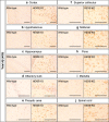
Figure 1.
Figure 1.
Widespread intraneuronal 4G8 immunoreactivity in the brains of HEXB KO mice. Coronal brain sections of HEXB KO mice were stained with 4G8 antibody, which recognizes amino acids 17–24 of Aβ and detects Aβ, full-length APP and APP-CTFs. Intraneuronal 4G8 signals were observed in most brain regions, including the cortex (a), hypothalamus (b), hippocampus (c), olfactory bulb (d), preoptic area (e), superior colliculus (f), midbrain (g), pons (h), medulla (i), and spinal cord (j). Scale bars: a–j, 200 μm.
Figure 2.
Figure 2.
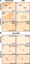
Accumulation of α-synuclein and pTau in HEXB KO mice. A, Brain sections of HEXB KO mice were costained with anti-α-synuclein (a, b) and 4G8 anti-Aβ (c, d) antibodies. α-Synuclein-LIR was detected in the SNC (b, b′), whereas intraneuronal 4G8 signals were detected in the SNR (d, d′). B, Immunohistochemical analysis of wild-type and HEXB KO mice using an antibody (AT8) that detects Tau phosphorylated at serine 202 and threonine 205 shows that pTau accumulates in the gray matter of the spinal cord (b) and the medulla (d) of HEXB KO mice. Arrows point to intraneuronal signals. Scale bars: a–d, 500 μm; b′, d′, 200 μm.
Figure 3.
Figure 3.
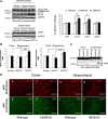
Accumulation of APP-CTFs and lipid-associated Aβ in HEXB KO mice. A, Western blot analysis, using 369 antibody to detect full-length APP and APP-CTFs and 22C11 antibody to detect sAPPα, shows significant increases in the levels of α-CTF and β-CTF in HEXB KO mice relative to wild-type (WT). No significant changes were observed in the levels of full-length APP or sAPPα levels. (For APP, α- and β-CTF analysis, n = 6 for WT, n = 7 for HZ, n = 7 for KO. For sAPPα, n = 3 for WT, n = 4 for HZ, n = 4 for KO). B, ELISA measurement of Aβ40 and Aβ42 levels shows significant increase in the levels of lipid-associated Aβ40 and Aβ42 in the HEXB KO mice (n = 6 for WT; n = 7 for HZ; n = 7 for KO). Shown are mean ± SEM (*p < 0.05; **p < 0.01; ***p < 0.001). C, Western blot analysis using Aβ40-specific antibody (FCA3340) shows increase in the levels of Aβ40 in the soluble and lipid-associated fractions of HEXB KO mice. The levels of Aβ42 were not detectable by Western blotting. D, Brain sections were costained with antibodies that recognize either the N terminus (22C11) or C terminus (369) of APP. Although the signals from APP-C terminus (369) antibody seem to accumulate in the cortex (d) and hippocampus (h) of HEXB KO mice, no change is observed in the signals from APP-N terminus (22C11) antibody (c, g). Scale bars: a–h, 200 μm.
Figure 4.
Figure 4.
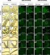
Region-specific accumulation of Aβ-like immunoreactivity in HEXB KO mice. Coronal brain sections of wild-type and HEXB KO mice were stained with NeuN antibody (a–j), Aβ42-specific antibody (FCA3542) (a1–j1), or Aβ40-specific antibody (FCA3340) (a2–j2). NeuN-positive neurons appeared swollen with vacuolar changes in the cortex (b, b′), subiculum (d, d′), dentate gyrus (f), and CA3 region of the hippocampus (h) of HEXB KO mice. Similarly, iAβ-LIR accumulated in the cortex (b1, b1′, b2, b2′), subiculum (d1, d1′, d2, d2′), dentate gyrus (f1, f2), and CA3 region of the hippocampus (h1, h2). The morphology of NeuN-positive neurons appeared normal in the CA1 region of the hippocampus (j), and iAβ-LIR was not observed in this region (j1, j2). DG, Dentate gyrus. Scale bars: a–j, a1–d1, g1–j1, a2–d2, g2–j2, 500 μm; a′–d′, a1′–d1′, a2′–d2′, e1, f1, e2, f2, 200 μm.
Figure 5.
Figure 5.
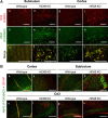
Ganglioside-bound Aβ (GAβ) immunoreactivity colocalizes with iAβ-LIR. A, Coronal brain sections from wild-type (a–c, g–i) and HEXB KO mice (d–f, j–l) were costained with GAβ antibody (4396C) and Aβ42-specific antibody (FCA3542). GAβ immunoreactivity as detected by 4396C antibody colocalized with iAβ-LIR in the subiculum and cortex of HEXB KO mice (f, l). Scale bars: a–l, 200 μm. B, Coronal brain sections from wild-type and HEXB KO mice were costained with GFAP (red)- and Aβ40 and Aβ42 (green)-specific antibodies (FCA3340 and FCA3542, respectively). Increased GFAP immunoreactivity and reactive gliosis were observed in the cortex (b), subiculum (d), and CA3 region of the hippocampus (f, h) of HEXB KO mice. Scale bars: a–h, 500 μm.
Figure 6.
Figure 6.
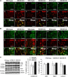
Dysfunction in lysosomal proteolysis leads to the accumulation of iAβ-LIR in the endosomal–lysosomal system and undegraded autophagosomes. A, Coronal brain sections of wild-type and HEXB KO mice were costained with Aβ42-specific antibodies (FCA3542) and markers of early endosomes (EEA1), late endosomes (Rab7), and lysosomes (Lamp-1). iAβ-LIR colocalized partially with EEA1 (a–f), Rab7 (g–l), and Lamp-1 (m–r) in HEXB KO mice. Images were taken from the subiculum. Scale bars, 50 μm. B, Immunohistochemical analysis shows the colocalization of iAβ42 (Pan1G6) with LC3 in undegraded autophagosomes in HEXB KO mice (d–f). The autophagosomal marker (LC3) colocalizes with the early endosomal marker (EEA1) (j–l) and with the lysosomal marker (Lamp-1) (p–r) in HEXB KO mice. Scale bars, 50 μm. C, Western blot analysis shows an increase in the LC3II/LC3I ratio and slight increases in the levels of p62 and Rab7 in HEXB KO mice relative to wild type (n = 3 for WT; n = 3 for HZ; n = 3 for KO). Shown are mean ± SEM.
Figure 7.
Figure 7.
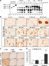
Human Sandhoff brains accumulate intraneuronal 4G8 immunoreactivity and APP-CTFs. A, Thin-layer chromatography analysis of cortices derived from GM1 gangliosidosis (GM1), SD, TS, AD, and age-matched controls, shows the levels of different gangliosides in each brain sample. B, GM1, SD, TS, AD, and age-matched control-derived cortical sections were immunostained using the monoclonal antibody 4G8. The arrows indicate accumulation of intraneuronal APP/Aβ-LIR in 1-year-old (e, f) and 2-year-old (g) SD brains; 1-year-old (h) and 19-year-old (i) GM1 brains; 4-year-old (j), 27-year-old (k), and 45-year-old (l) TS brains. Scale bars: a–l, 200 μm. C, The specificity of 4G8 staining was confirmed by immunostaining cortical sections derived from human SD and control brains with 4G8 antibody preincubated with 100× molar excess of Aβ1–40 peptide (b, e), or with secondary antibody only (c, f). Scale bars: a–f, 200 μm. D, Western blot analysis of seven fibroblast cell lines derived from SD patients or healthy controls show significant accumulation of α-CTFs in the SD fibroblasts (**p = 0.007). Values are the mean of three individual experiments. Error bars indicate SEM.
Figure 8.
Figure 8.
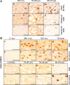
Human gangliosidoses brains accumulate intraneuronal ganglioside-bound Aβ. A, Immunostaining of human brain cortical sections using the antibodies 4G8, FC3542 (Aβ42-specific antibody), and FC3340 (Aβ40-specific antibody) demonstrates that Aβ42 accumulates in SD (b) and TS brains (e). Extracellular Aβ-like immunoreactivity structures were observed in the 45-year-old TS brain (e). The arrows indicate accumulation of intraneuronal Aβ-LIR. Scale bars: a–f, 200 μm; g–i, 500 μm. B, Cortical sections obtained from control (1 year) (a, b), control (27 years) (i, j), GM1 (1 year) (c, d), SD (1 year) (e, f), TS (4 years) (g, h), TS (27 years) (k, l), TS (45 years) (m, n), and AD (75 years) (o, p) were stained with 4396C antibody to detect ganglioside-bound Aβ. Scale bars: a, b, o, p, 200 μm; c–n, 50 μm.

Similar articles

Cited by

References

    1. Ariga T, McDonald MP, Yu RK. Role of ganglioside metabolism in the pathogenesis of Alzheimer's disease—a review. J Lipid Res. 2008;49:1157–1175. - PMC - PubMed
    1. Bahr BA, Bendiske J. The neuropathogenic contributions of lysosomal dysfunction. J Neurochem. 2002;83:481–489. - PubMed
    1. Barelli H, Lebeau A, Vizzavona J, Delaere P, Chevallier N, Drouot C, Marambaud P, Ancolio K, Buxbaum JD, Khorkova O, Heroux J, Sahasrabudhe S, Martinez J, Warter JM, Mohr M, Checler F. Characterization of new polyclonal antibodies specific for 40 and 42 amino acid-long amyloid beta peptides: their use to examine the cell biology of presenilins and the immunohistochemistry of sporadic Alzheimer's disease and cerebral amyloid angiopathy cases. Mol Med. 1997;3:695–707. - PMC - PubMed
    1. Boland B, Kumar A, Lee S, Platt FM, Wegiel J, Yu WH, Nixon RA. Autophagy induction and autophagosome clearance in neurons: relationship to autophagic pathology in Alzheimer's disease. J Neurosci. 2008;28:6926–6937. - PMC - PubMed
    1. Boland B, Smith DA, Mooney D, Jung SS, Walsh DM, Platt FM. Macroautophagy is not directly involved in the metabolism of amyloid precursor protein. J Biol Chem. 2010;285:37415–37426. - PMC - PubMed

Publication types

MeSH terms