. 2012 Apr 24;109(17):6757-62.
doi: 10.1073/pnas.1118814109. Epub 2012 Apr 10.

Spontaneous spatiotemporal waves of gene expression from biological clocks in the leaf

Affiliations

Spontaneous spatiotemporal waves of gene expression from biological clocks in the leaf

Bénédicte Wenden et al. Proc Natl Acad Sci U S A. .

Abstract

The circadian clocks that drive daily rhythms in animals are tightly coupled among the cells of some tissues. The coupling profoundly affects cellular rhythmicity and is central to contemporary understanding of circadian physiology and behavior. In contrast, studies of the clock in plant cells have largely ignored intercellular coupling, which is reported to be very weak or absent. We used luciferase reporter gene imaging to monitor circadian rhythms in leaves of Arabidopsis thaliana plants, achieving resolution close to the cellular level. Leaves grown without environmental cycles for up to 3 wk reproducibly showed spatiotemporal waves of gene expression consistent with intercellular coupling, using several reporter genes. Within individual leaves, different regions differed in phase by up to 17 h. A broad range of patterns was observed among leaves, rather than a common spatial distribution of circadian properties. Leaves exposed to light-dark cycles always had fully synchronized rhythms, which could desynchronize rapidly. After 4 d in constant light, some leaves were as desynchronized as leaves grown without any rhythmic input. Applying light-dark cycles to such a leaf resulted in full synchronization within 2-4 d. Thus, the rhythms of all cells were coupled to external light-dark cycles far more strongly than the cellular clocks were coupled to each other. Spontaneous desynchronization under constant conditions was limited, consistent with weak intercellular coupling among heterogeneous clocks. Both the weakness of coupling and the heterogeneity among cells are relevant to interpret molecular studies and to understand the physiological functions of the plant circadian clock.


Conflict of interest statement

The authors declare no conflict of interest.

Figures

Fig. 1.
Fig. 1.
Setup for imaging luciferase in intact and detached leaves over several days. (A) Setup for one detached leaf; (B) red and blue light-emitting diode (LED) system for intact plant imaging under the microscope; (C) luminescence output for four six-well plates as imaged in a cabinet; (D) luminescence output in pseudocolor from a CCA1:LUC leaf imaged (25×) as in B. Movie S1 shows the time series from a similar leaf.
Fig. 2.
Fig. 2.
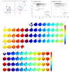
Spatiotemporal analysis of CCA1:LUC rhythms in an intact leaf entrained under light–dark cycles and imaged under constant light. Plants were entrained under LD 12:12 cycles for 12 d and then transferred and imaged under LL conditions. (A) Average luminescence, detrended, for a CCA1:LUC leaf (Table S1, plant 23); (B) detrended luminescence for all pixels of the leaf in A; (C) montage showing the spatial pattern of the luminescence in B (interval between images = 2 h); (D) data in C represented as a montage of the circadian phase (in radians) at each pixel; (E) phase at ZT48 along two central lines. The phase values are at the location of the pixel values, as shown by the overlaid lines. No colored square is created for the uppermost data points, at the leaf petiole. Arrow in C and D indicates the petiole. Time is in hours; ZT0 corresponds to transfer to LL.
Fig. 3.
Fig. 3.
Quantification of phase coherence and montages. (A) Calculation of phase coherence, R, of the phase vectors projected on the unit circle. (Left) At a particular time point, three pixels have phase angles θ1, θ2, and θ3. Addition of the vectors and division by n = 3 gives the mean resultant length, R. In this example R is close to 1, showing that the phases of the three pixels are rather tightly clustered. (Right) ϕ indicates the mean phase at this time point. (B) R values for intact plants and detached leaves. Red and green lines represent plant 3 and plant 16, respectively (Table S1). (C) Phase montage for a CCA1:LUC detached leaf, grown in LD for 21 d and imaged in LL (Table S1, plant 3); one cycle is 28 h. (D) Phase montage for a CCA1:LUC detached leaf, grown in LL for 21 d and imaged in LL (Table S1, plant 16); one cycle is 24 h. Interval between two images = 2 h. Time is in hours; ZT0 corresponds to the start of imaging. Arrow in C and D indicates the position of the petiole.
Fig. 4.
Fig. 4.
Spatiotemporal patterns in plants grown and imaged under constant light. (A and B) Peak firing patterns for two independent CCA1:LUC detached leaves (Table S1, plants 16 and 20, respectively) grown and imaged in LL. Black dots represent the leaf areas peaking at the time of the picture. Interval between two images = 40 min. (C and D) Mean period for two independent CCA1:LUC intact leaves (Table S1, plants 56 and 58, respectively), grown and imaged in LL. (E and F) Mean period for the detached leaves shown in A and B. Plants were grown in LL for 21 d (A and B) or 12 d (C and D) before imaging.
Fig. 5.
Fig. 5.
Resynchronization of nonentrained leaves. Plant 31 (Table S1) was grown under LL conditions for 13 d and transferred into LL and then LD for imaging. (A) Luminescence for all pixels, detrended for the CCA1:LUC leaf. (B) Montage of the detrended luminescence, interval between two images = 2 h. Time is in hours; ZT0 corresponds to start of imaging. Arrow in B indicates the position of the petiole.

References

    1. McWatters HG, Devlin PF. Timing in plants—a rhythmic arrangement. FEBS Lett. 2011;585:1474–1484. - PubMed
    1. Pokhilko A, et al. The clock gene circuit in Arabidopsis includes a repressilator with additional feedback loops. Mol Syst Biol. 2012;8:574. - PMC - PubMed
    1. Millar AJ, Short SR, Chua NH, Kay SA. A novel circadian phenotype based on firefly luciferase expression in transgenic plants. Plant Cell. 1992;4:1075–1087. - PMC - PubMed
    1. Plautz JD, Kaneko M, Hall JC, Kay SA. Independent photoreceptive circadian clocks throughout Drosophila. Science. 1997;278:1632–1635. - PubMed
    1. Thain SC, Hall A, Millar AJ. Functional independence of circadian clocks that regulate plant gene expression. Curr Biol. 2000;10:951–956. - PubMed

Publication types

MeSH terms