. 2012:2012:194723.
doi: 10.1155/2012/194723. Epub 2012 Feb 14.

Involvement of the ubiquitin-proteasome system in the formation of experimental postsurgical peritoneal adhesions

Affiliations

Involvement of the ubiquitin-proteasome system in the formation of experimental postsurgical peritoneal adhesions

Clara Di Filippo et al. Mediators Inflamm. 2012.

Abstract

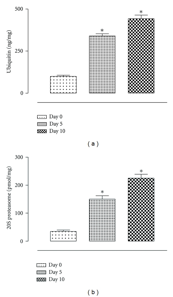
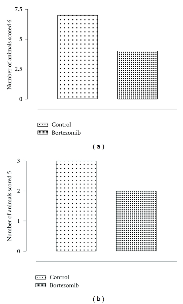
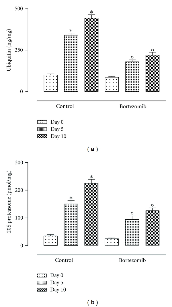
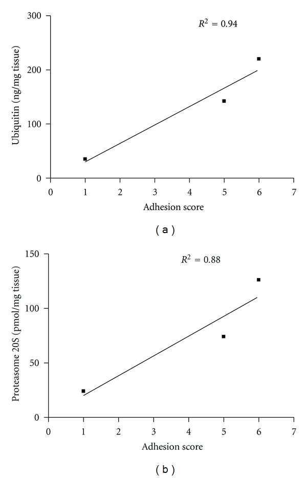
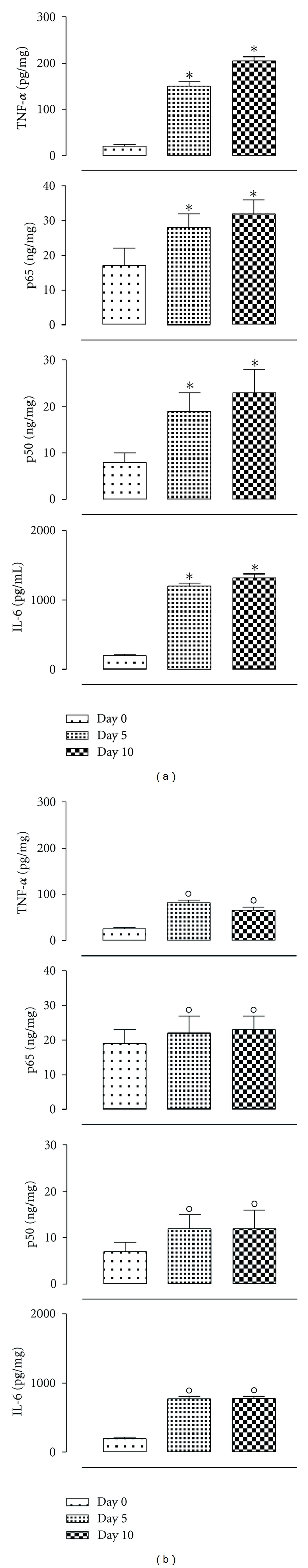
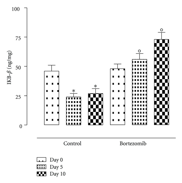
We investigated the Ubiquitin-Proteasome System (UPS), major nonlysosomal intracellular protein degradation system, in the genesis of experimental postsurgical peritoneal adhesions. We assayed the levels of UPS within the adhered tissue along with the development of peritoneal adhesions and used the specific UPS inhibitor bortezomib in order to assess the effect of the UPS blockade on the peritoneal adhesions. We found a number of severe postsurgical peritoneal adhesions at day 5 after surgery increasing until day 10. In the adhered tissue an increased values of ubiquitin and the 20S proteasome subunit, NFkB, IL-6, TNF-α and decreased values of IkB-beta were found. In contrast, bortezomib-treated rats showed a decreased number of peritoneal adhesions, decreased values of ubiquitin and the 20S proteasome, NFkB, IL-6, TNF-α, and increased levels of IkB-beta in the adhered peritoneal tissue. The UPS system, therefore, is primarily involved in the formation of post-surgical peritoneal adhesions in rats.


Figures

Figure 1
Figure 1
Levels of ubiquitin and 20S proteasome in the tissues from rats during peritoneal adhesions development at time 0, 5, and 10 days from the surgery in the control group. The differences from day 0 are considered with *P < 0.01.
Figure 2
Figure 2
Quantitative evaluation by the peritoneal adhesions score indicating a substantial decrease of adhesions in the group of rats treated with bortezomib after 10 days of surgery if compared with the control group same day.
Figure 3
Figure 3
Levels (ng/mg tissue) of ubiquitin and 20S proteasome in the tissues from rats during peritoneal adhesions development. At time 0, 5, and 10 in the group treated with bortezomib by intravenous injection (0.05 mg/kg) given 1 hour before surgery, a second injection was given 1 hour after the surgical procedure (total dose of 0.1 mg/kg) and in the control group. The differences from day 0 are considered with *P < 0.01, and the differences from the control group at the same day are considered as °P < 0.01.
Figure 4
Figure 4
Correlation between the adhesion score and the local levels of ubiquitin and protesome 20S 10 days after the surgery.
Figure 5
Figure 5
Levels (ng/mg tissue) of TNF-alpha, p50, p65, and IL-6 within the tissues from rats during peritoneal adhesions development at time 0, 5, and 10 days from the peritoneal surgery. The rats were treated or not with bortezomib by intravenous injection (0.05 mg/kg) given 1 hour before surgery, and a second injection was given 1 hour after the surgical procedure (total dose of 0.1 mg/kg). The differences from day 0 are considered with *P < 0.01, and the differences from the control group at the same day are considered as °P < 0.01.
Figure 6
Figure 6
IKB-β levels (ng/mg tissue) within the tissues from rats during peritoneal adhesions development. Rats at time 0, 5, and 10 days from the surgery and treated or not with bortezomib. The differences from day 0 are considered with *P < 0.01, and the differences from the control group at the same day are considered as °P < 0.01.

Similar articles

Cited by

References

    1. Canis M, Botchorishvili R, Wattiez A, et al. Prevention of peritoneal adherences. Journal de Gynecologie Obstetrique et Biologie de la Reproduction. 2001;30(4):305–324. - PubMed
    1. Attard JAP, Maclean AR. Adhesive small bowel obstruction: epidemiology, biology and prevention. Canadian Journal of Surgery. 2007;50(4):291–300. - PMC - PubMed
    1. Ott DE. Laparoscopy and adhesion formation, adhesions and laparoscopy. Seminars in Reproductive Medicine. 2008;26(4):322–330. - PubMed
    1. Di Filippo C, Falsetto A, De Pascale V, et al. Plasma levels of t-PA and PAI-1 correlate with the formation of experimental post-surgical peritoneal adhesions. Mediators of Inflammation. 2006;2006:4 pages. Article ID 13901. - PMC - PubMed
    1. Lauder CIW, Garcea G, Strickland A, Maddern GJ. Abdominal adhesion prevention: still a sticky subject? Digestive Surgery. 2010;27(5):347–358. - PubMed

Publication types