. 2012 Mar;14(3):178-89.
doi: 10.1593/neo.111444.

IκB kinases modulate the activity of the androgen receptor in prostate carcinoma cell lines

Affiliations

IκB kinases modulate the activity of the androgen receptor in prostate carcinoma cell lines

Garima Jain et al. Neoplasia. 2012 Mar.

Abstract

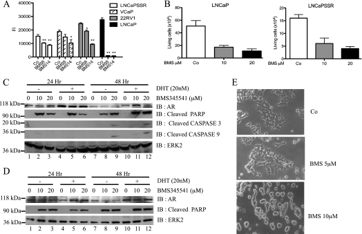
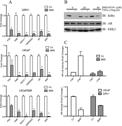
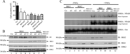
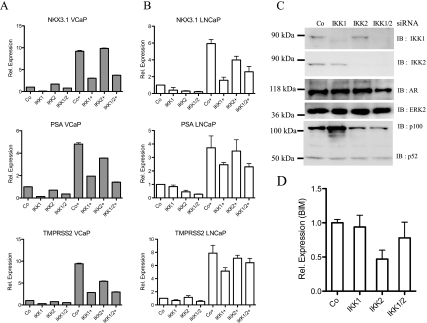
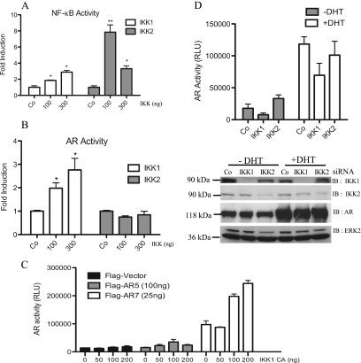
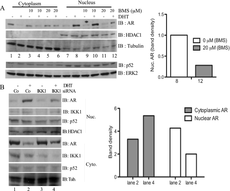
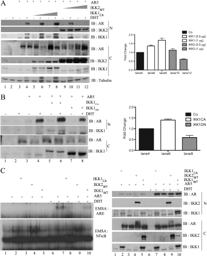
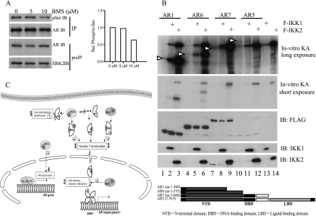
Enhanced nuclear localization of nuclear factor κB (NF-κB) in prostate cancer (PCa) samples and constitutive NF-κB signaling in a class of PCa cell lines with low androgen receptor (AR) expression (PC3 and DU-145) imply an important role of the IκB kinase (IKK)/NF-κB system in PCa. However, most PCa and PCa cell lines depend on the activity of the AR, and the role of NF-κB in these AR-expressing PCa remains unclear. Here, we demonstrate that inhibition of NF-κB signaling by the IKK inhibitor BMS345541 reduced proliferation and increased apoptosis in AR-expressing PCa cell lines. Furthermore, AR activity and target gene expression were distinctively reduced, whereas AR protein levels remained unaltered on BMS345541 treatment. Similar effects were observed particularly after small interfering RNA (siRNA)-mediated knockdown of IKK1, but not by siRNA-mediated suppression of IKK2. Moreover, IKK1 overexpression augmented 5α-dihydrotestosterone-induced nuclear AR translocation, whereas nuclear AR was reduced by IKK1 knockdown or BMS345541. However, because IKK1 also enhances the activity of a chronically nuclear AR mutant, modulation of the subcellular distribution seems not to be the only mechanism by which IKK1 enhances AR activity. Finally, reduced in vivo AR phosphorylation after BMS345541 treatment and in vitro AR phosphorylation by IKK1 or IKK2 imply that AR constitutes a novel IKK target. Taken together, our data identify IKK1 as a potentially target structure for future therapeutic intervention in PCa.


Figures

Figure 1
Figure 1
Growth inhibition of androgen-dependent and androgen-independent PCa cells by BMS345541. (A) Cells were either left untreated or treated with BMS345541 (8 µM; 14 µM) for 24 hours, and cell proliferation was measured by reduction of AlamarBlue dye (bars depict mean ± SD, **P < .0001, *P < .001, compared with vehicle control). (B) Twenty-four hours after exposure to BMS345541 (0, 10, and 20 µM) of LNCaP and LNCaP-SSR cells, viable cell numbers were determined. Graph indicates mean ± SD. LNCaP (C) and LNCaP-SSR (D) cells were treated with BMS345541 for 24 and 48 hours. Cells were left unstimulated (-) or stimulated with 20 nM DHT (+), and whole-cell extracts were subjected to immunoblot analysis with the indicated antibodies. (E) LNCaP cells were either left untreated (Co) or were treated with indicated concentrations of BMS345541 for 24 hours. Cell morphology is depicted as bright field images (original magnification, x20).
Figure 2
Figure 2
BMS345541 inhibits transcriptional activity of AR and NF-κB. (A) LNCaP, LNCaP-SSR, and 22Rv1 cells were either left untreated (Co) or treated with 8 µM BMS345541 for 24 hours (BMS). Quantitative PCR analysis of AR, AR target genes (PSA, PME, NKX3.1, and TMPRSS2), and NF-κB target gene (BIM) was performed. Normalized readings were plotted relative to Co of each gene. Bar depicts mean ± SD. *P < .001, **P < .0001. (B) LNCaP cells were pretreated for 4 hours with BMS345541 (0, 20, and 40 µM) followed by TNFα stimulation for 20 minutes. Resulting whole-cell extracts were subjected to immunoblot analysis for the indicated proteins. (C) LNCaP cells were transiently transfected with a 3xκB-luc (upper part) or with the MMTVluc (lower part) and treated with BMS345541 (8 µM) for 24 hours. Cells transfected with 3xκB-luc were additionally stimulated with TNFα (+TNF) or were not further stimulated (-TNF) for 3 more hours. Subsequently, dual luciferase analysis was performed with all samples. Bars depict mean ± SD of normalized luciferase reading relative to untreated samples.
Figure 3
Figure 3
Effect of siRNA-mediated suppression of IKK1 and/or IKK2. (A) LNCaP cells were transfected with the indicated IKK siRNA. Four days after transfection, viable cell numbers were determined and plotted as mean ± SD. (B) Efficiency of knockdown corresponding to A was checked by immunoblot analysis. The same membrane was reprobed with antibodies recognizing AR and ERK2. (C) LNCaP cells were transfected with IKK1 and/or IKK2 siRNA, as indicated. After 72 hours, cells either were left untreated (-TNFα) or were stimulated with 10 ng/ml TNFα for 20 minutes. EMSA for NF-κB or Oct probes was performed with nuclear extract of these cells (upper panel). Corresponding cytoplasmic extracts were used for immunoblot analysis with indicated antibodies (lower panel).
Figure 4
Figure 4
IKK1 siRNA attenuates the expression of AR target genes. VCaP (A) and LNCaP cells (B) were transfected with IKK1 and/or IKK2 siRNA. Forty-eight hours after transfection, cells were either left untreated or treated with 20 nM DHT (+) for an additional 24 hours. Transcript levels of AR, PSA, NKX3.1, and TMPRSS2 were analyzed with real-time PCR. Histogram depicts fold change in transcript levels after siRNA transfection relative to Co-siRNA-transfected samples. (C) IKK1, IKK2, and AR protein levels in corresponding LNCaP samples were determined with immunoblot analysis. (D) Similar quantitative PCR analysis of BIM transcript levels after knockdown of IKK1 and/or IKK2 siRNA in LNCaP cells.
Figure 5
Figure 5
Effects of IKK1 and IKK2 on AR activity. (A, B) LNCaP cells were transfected with IKK1CA or IKK2WT (0, 100, and 300 ng), along with either 3xκB-luc (A) or MMTVluc (B). After 24 hours, dual luciferase analysis was performed. Bars in histogram represent mean ± SD of normalized relative light unit (RLU) relative to IKK-untransfected samples. *P ≤ .03, **P ≤ .001, compared with control always. (C) COS7 cells were transfected with MMTVluc and Ubi-Renilla and with increasing amounts of IKK1CA along with either full-length AR (AR5) or constitutive active AR (AR7). Luciferase values were determined 24 hours after transfection. The mean values ± SD of the normalized RLU are depicted. (D) 293 cells were cotransfected with IKK siRNA along with MMTVluc and Ubi-Renilla. Twenty-four hours after transfection, cells were stimulated with 20 nM DHT (+DHT) or left untreated (-DHT) for another 24 hours. Mean ± SD of normalized RLU value of respective transfection is depicted (upper part). Knockdown efficiency was controlled by immunoblot analysis with the corresponding samples (lower part).
Figure 6
Figure 6
Effect of IKK inhibition and IKK1 knockdown on nuclear translocation of the AR. (A) LNCaP cells were cultivated in Dulbecco modified Eagle medium (DMEM) + charcoal-stripped fetal calf serum (CS-FCS) for 2 hours before pretreatment with BMS345541 (0, 10, and 20 µM) for an additional 1 hour. Subsequently, cells were either left untreated (-) or treated (+) with DHT for 1 hour, and nuclear and cytoplasmic proteins were subjected to immunoblot analysis using anti-AR, anti-HDAC1, and anti-tubulin antibodies. The subcellular distribution of p52 was determined with immunoblot analysis in a similar experiment (lower part). (B) LNCaP cells were transiently transfected with either control siRNA (Co) or with IKK1si2 siRNA (IKK1). After 48 hours, the cells were transferred to DMEM + CS-FCS for 3 hours followed by treatment with either vehicle (DMSO, -) or with DHT (5 nM, +) for an additional hour. Subsequently, nuclear and cytoplasmic proteins were extracted and subjected to immunoblot analyses with antibodies for the indicated proteins. A quantification of the cytoplasmic and the nuclear AR protein levels in DHT-stimulated cells (lanes 2 and 4) is given in the right part.
Figure 7
Figure 7
Differential effects of IKK1 or IKK2 coexpression on nuclear expression and DNA binding of the AR. (A) COS7 cells expressing ectopic AR5 alone or in combination with FLAG-IKK1CA or FLAG-IKK2WT as indicated were either left untreated (-) or stimulated with 20 nM DHT (+) for 1 hour. The resulting nuclear and cytoplasmic extracts were subjected to immunoblot analysis. This experiment was repeated twice, and a quantification of selected samples of all three experiments is given in the right part. (B) COS7 cells, transiently transfected with FLAG-AR5 alone or with FLAG-IKK1CA or HA-IKK1DN, were incubated in DMEM + CSS before stimulation with DHT for 1 hour. Nuclear and cytoplasmic extracts were subjected to immunoblot analysis with the indicated antibodies. This experiment was repeated twice, and a quantification of selected samples of all three experiments is given in the right part. (C) COS7 were transiently transfected with FLAG-AR5, FLAG-IKK1CA, FLAG-IKK2WT, and FLAG-IKK1WT as indicated. Subsequently, cells were treated either with solvent or with DHT (+) for 1 hour. Nuclear extracts were subjected to EMSA experiments using NF-κB (left part, lower panel) or AR (left part, upper panel) specific oligonucleotides. In addition, immunoblot analysis with the indicated antibodies was performed with the cytoplasmic (C) and nuclear (N) proteins (right part).
Figure 8
Figure 8
The AR is an IKK target in vitro. (A) LNCaP cells were exposed to BMS345541 (0, 5, and 10 µM). After 48 hours, AR was immunoprecipitated (IP), and anti-phospho-serine, AR, and ERK2 immunoblot analysis was performed. Histogram depicts densitometry of anti-phospho-serine signals relative to control. (B) 293 cells expressing AR5, AR deletion mutants, IKK1, or IKK2 were lysed, and anti-FLAG immunoprecipitation was performed. Kinase reaction was performed with AR proteins alone or with either IKK1 or IKK2 and analyzed with autoradiography. Arrows indicate the position of AR variants. A schematic representation of AR variants is given in the lower part. (C) Model depicting the mechanism by which the IKK proteins control the activity of the AR: DHT binding induced nuclear localization, ARE binding, and target gene induction by AR. IKK1 phosphorylates AR in the nucleus and/or cytoplasm causing an increased nuclear localization of AR (1). IKK1 additionally affects transcriptional activity of AR independent of its nuclear localization possibly by altered DNA binding ability or Cofactor interaction (2). IKK2 attenuates ligand-dependent nuclear localization of AR most likely by phosphorylating AR at alternative sites (3). Conversely, IKK2 regulates AR expression (4). In addition, IKK2 seems to affect cytoplasmic AR by modifying AR after translation (5).

Similar articles

Cited by

References

    1. Linja MJ, Savinainen KJ, Saramaki OR, Tammela TL, Vessella RL, Visakorpi T. Amplification and overexpression of androgen receptor gene in hormone-refractory prostate cancer. Cancer Res. 2001;61:3550–3555. - PubMed
    1. Zhao XY, Malloy PJ, Krishnan AV, Swami S, Navone NM, Peehl DM, Feldman D. Glucocorticoids can promote androgen-independent growth of prostate cancer cells through a mutated androgen receptor. Nat Med. 2000;6:703–706. - PubMed
    1. Schutz SV, Schrader AJ, Zengerling F, Genze F, Cronauer MV, Schrader M. Inhibition of glycogen synthase kinase-3β counteracts ligand-independent activity of the androgen receptor in castration resistant prostate cancer. PLoS One. 2011;6:e25341. - PMC - PubMed
    1. Oeckinghaus A, Ghosh S. The NF-κB family of transcription factors and its regulation. Cold Spring Harb Perspect Biol. 2009;1:a000034. - PMC - PubMed
    1. Scheidereit C. IκB kinase complexes: gateways to NF-κB activation and transcription. Oncogene. 2006;25:6685–6705. - PubMed

Publication types