

doi: 10.1371/journal.pcbi.1002445.
Epub 2012 Mar 29.
Circular permutation in proteins
Affiliations
- PMID: 22496628
- PMCID: PMC3320104
- DOI: 10.1371/journal.pcbi.1002445
Item in Clipboard
Circular permutation in proteins
PLoS Comput Biol.
2012.
No abstract available
Conflict of interest statement
The authors have declared that no competing interests exist.
Figures
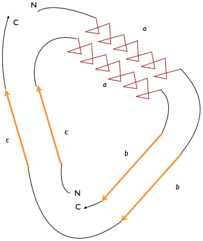
The first protein (outer circle) has the sequence a-b-c. After the permutation the second protein (inner circle) has the sequence c-a-b. The letters N and C indicate the location of the amino- and carboxy-termini of the protein sequences and how their positions change relative to each other.
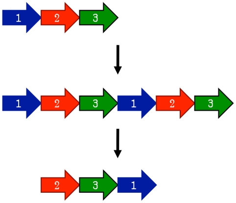
First, a gene is duplicated in place. Next, start and stop codons are introduced, resulting in a circularly permuted gene.
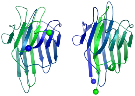
They could have evolved from a similar gene . Both consist of four alpha helices with the order of helices being permuted relative to each other.
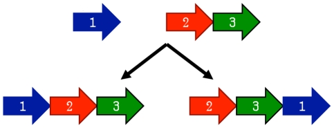
Two separate genes arise (potentially from the fission of a single gene). If the genes fuse together in different orders in two orthologues, a circular permutation occurs.
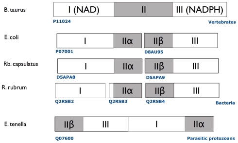
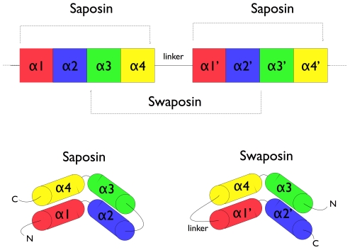
In cattle, the three domains are arranged sequentially. In the bacteria E. coli, Rb. capsulatus, and R. rubrum, the transhydrogenase consists of two or three subunits. Finally, transhydrogenase from the protist E. tenella consists of a single subunit that is circularly permuted relative to cattle transhydrogenase .
References
-
- Einspahr H, Parks EH, Suguna K, Subramanian E, Suddath FL. The crystal structure of pea lectin at 3.0-A resolution. J Biol Chem. 1986;261:16518–16527. - PubMed
-
- Carrington DM, Auffret A, Hanke DE. Polypeptide ligation occurs during post-translational modification of concanavalin A. Nature. 1985;313:64–67. - PubMed
-
- Bowles DJ, Pappin DJ. Traffic and assembly of concanavalin A. Trends Biochem Sci. 1988;13:60–64. - PubMed
-
- Goldenberg DP, Creighton TE. Circular and circularly permuted forms of bovine pancreatic trypsin inhibitor. J Mol Biol. 1983;165:407–413. - PubMed
Publication types
MeSH terms
Substances
LinkOut - more resources
Full Text Sources
Other Literature Sources
