. 2012;8(4):e1002627.
doi: 10.1371/journal.ppat.1002627. Epub 2012 Apr 5.

Small-molecule inhibitors of dengue-virus entry

Affiliations

Small-molecule inhibitors of dengue-virus entry

Aaron G Schmidt et al. PLoS Pathog. 2012.

Erratum in

Abstract

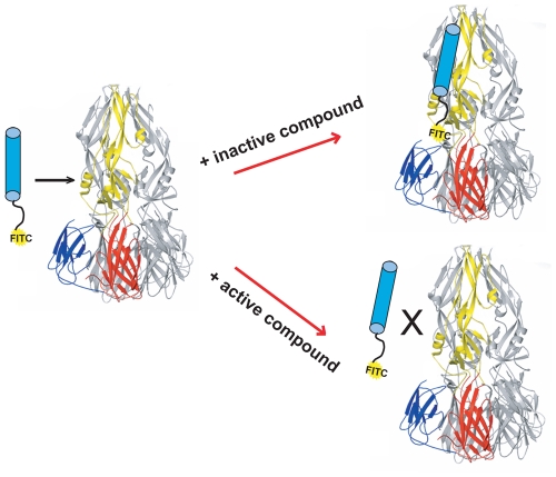
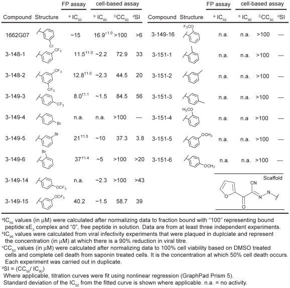
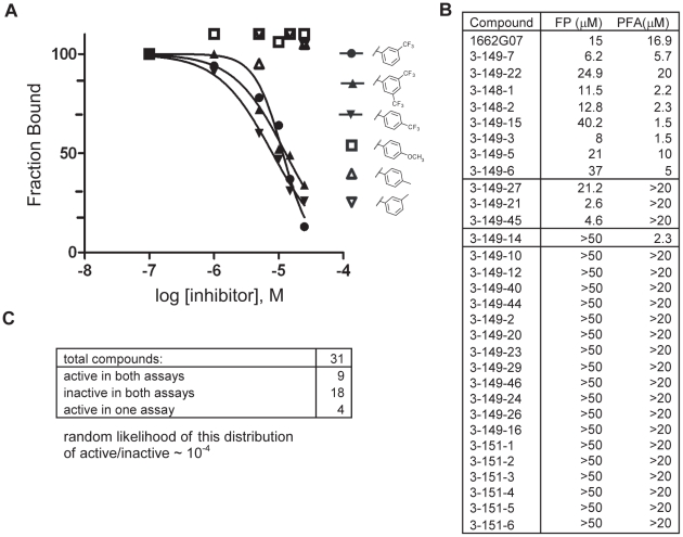
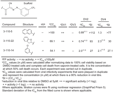
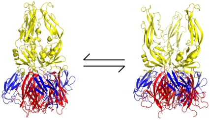
Flavivirus envelope protein (E) mediates membrane fusion and viral entry from endosomes. A low-pH induced, dimer-to-trimer rearrangement and reconfiguration of the membrane-proximal "stem" of the E ectodomain draw together the viral and cellular membranes. We found stem-derived peptides from dengue virus (DV) bind stem-less E trimer and mimic the stem-reconfiguration step in the fusion pathway. We adapted this experiment as a high-throughput screen for small molecules that block peptide binding and thus may inhibit viral entry. A compound identified in this screen, 1662G07, and a number of its analogs reversibly inhibit DV infectivity. They do so by binding the prefusion, dimeric E on the virion surface, before adsorption to a cell. They also block viral fusion with liposomes. Structure-activity relationship studies have led to analogs with submicromolar IC₉₀s against DV2, and certain analogs are active against DV serotypes 1,2, and 4. The compounds do not inhibit the closely related Kunjin virus. We propose that they bind in a previously identified, E-protein pocket, exposed on the virion surface and although this pocket is closed in the postfusion trimer, its mouth is fully accessible. Examination of the E-trimer coordinates (PDB 1OK8) shows that conformational fluctuations around the hinge could open the pocket without dissociating the trimer or otherwise generating molecular collisions. We propose that compounds such as 1662G07 trap the sE trimer in a "pocket-open" state, which has lost affinity for the stem peptide and cannot support the final "zipping up" of the stem.


Conflict of interest statement

The authors have declared that no competing interests exist.

Figures

Figure 1
Figure 1. Schematic of high-throughput screening platform.
DV2 sE3 with one monomer colored by domain: DI (red), DII (yellow) and DIII (blue). Stem peptide DV2419–447, with FITC at its N-terminus, is represented as a cylinder, conjugated at its N-terminus with FITC. Addition of small molecules from screening libraries either affect (active compounds) or do not affect (inactive compounds) interaction of stem peptide with sE3.
Figure 2
Figure 2. Biochemical, cytotoxicity and antiviral summary of 1662G07 select compounds from the 3-148, 149 and 151 series.
Figure 3
Figure 3. Activity profiles of selected compounds in competition FP and PFA assays.
(A) Representative FP titration curves of active analogs: 3-148-1, 3-148-2 and 3-149-3 and inactive analogs: 3-151-2, 3-151-3 and 3-151-6 and their competition with the interaction between stem peptide and sE3. (B) Activities in the two assays for all compounds in the SAR series which varied at R2 (as phenyl). Table 1 and Supplementary Table 2 give the chemical structures of all compounds in this list. (C) Summary of the numbers of active and inactive compounds in each of the two assays. The likelihood that this degree of concordance could result from random pairing of unrelated activities is less than 10−4. Only 9 of the 31 compounds had activities within the range of achievable concentrations, making a quantitative correlation of those activities uninformative, particularly in few of other sources of error such as nonspecific binding to cell surfaces in the case of the infectivity assay and limited solubility of some compounds in the case of both assays.
Figure 4
Figure 4. Biochemical, cytotoxicity and antiviral summary of selected compounds from the 3-110 series.
Figure 5
Figure 5. Effect of order-of-addition on small-molecule inhibition.
(A) Comparison of o-, m-, and p-OCF3 and m-, di-m- and p-CF3 substitution from the 3-148 and 3-149 series (B) Comparison of compounds from the 3-110 series. Preincubation: addition of 1662G07 analogs to inoculum 15′ before adsorption to cells. Coinfection: addition of analogs at the time of adsorption. Postinfection: addition of analogs one hour after adsorption of virus. In all cases, cells were washed with PBS before adding compounds. Supernatants were harvested after 24 hours and viral titres determined by standard plaque forming assay (done in duplicate). Compounds from (A) and (B) were used at 15 and 5 µM, respectively. DV2419–447 stem peptide at 1 µM was used as a control.
Figure 6
Figure 6. Inhibition of viral fusion with liposomes.
Effect on content mixing of preincubating virus with 1662G07 analogs. Virus and analogs 3-148-1, 3-149-15, 3-110-5 and 3-110-22 (all at 50 µM) were incubated with liposomes encapsulating trypsin and acidified to pH=5.5. Following back-neutralization and incubation for 1 hr at 37 C, samples were prepared for SDS-PAGE and immunoblotted with αC and αE antibody. Fusion leads to exposure of core protein to trypsin and loss of the corresponding band but retention of the envelope protein band. DV2419–447 stem peptide, at 1 µM, was used as a positive control.
Figure 7
Figure 7. Interaction of 1662G07 analogs with DI/DII.
DI/DII was immobilized on a CM5 sensorchip. Analogs 3-148-1, 3-149-3, 3-149-14, 3-151-2, 3-151-2, 3-151-5, 3-151-4, 3-110-5, 3-110-14 and 3-110-22 were passed over the DI/DII surface at 10, 20 and 40 µM. Background for nonspecific binding to the chip surface was corrected for by passing the analogs over a protein-free channel. All measurements carried out in duplicate.
Figure 8
Figure 8. Reversibility of antiviral effect.
Viral inocula were preincubated with 1662G07 analogs from the (A) 3-148 and 3-149 and (B) 3-110 series for 10′ at 37°C. DI/DII was then added in molar excess and the incubation continued for an additional 15′. Each inoculum was added to cells, and supernatants were harvested 24 hrs later. An inoculum preincubated with DI/DII alone at the same molar excess showed no loss in viral titre.
Figure 9
Figure 9. Proposed mechanism of action of small-molecule inhibitors and postulated equilibrium between two conformations of the sE trimer.
In the “pocket-open", inhibitor-stabilized conformation (right image), the stem-binding groove is absent and the final fusion-inducing step in the conformational change cannot occur. Moreover, sE in this pocket-open conformation would not bind stem-derived peptides. Domains I, II and III are in red, yellow and blue, respectively. All images created with PyMol.

References

    1. Harrison SC. Mechanism of membrane fusion by viral envelope proteins. Adv Virus Res. 2005;64:231–261. - PMC - PubMed
    1. Harrison SC. Viral membrane fusion. Nat Struct Mol Biol. 2008;15:690–698. - PMC - PubMed
    1. van der Schaar HM, Rust MJ, Chen C, van der Ende-Metselaar H, Wilschut J, et al. Dissecting the cell entry pathway of dengue virus by single-particle tracking in living cells. PLoS Pathog. 2008;4:e1000244. - PMC - PubMed
    1. van der Schaar HM, Rust MJ, Waarts BL, van der Ende-Metselaar H, Kuhn RJ, et al. Characterization of the early events in dengue virus cell entry by biochemical assays and single-virus tracking. J Virol. 2007;81:12019–12028. - PMC - PubMed
    1. Fritz R, Stiasny K, Heinz FX. Identification of specific histidines as pH sensors in flavivirus membrane fusion. J Cell Biol. 2008;183:353–361. - PMC - PubMed

Publication types

MeSH terms