. 2012;8(4):e1002635.
doi: 10.1371/journal.ppat.1002635. Epub 2012 Apr 5.

Leishmania induces survival, proliferation and elevated cellular dNTP levels in human monocytes promoting acceleration of HIV co-infection

Affiliations

Leishmania induces survival, proliferation and elevated cellular dNTP levels in human monocytes promoting acceleration of HIV co-infection

David J Mock et al. PLoS Pathog. 2012.

Abstract

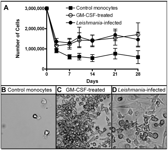
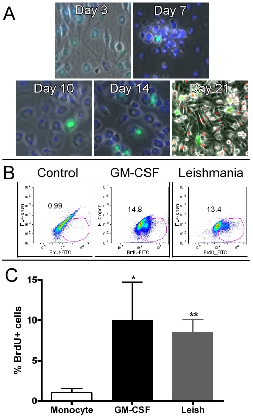
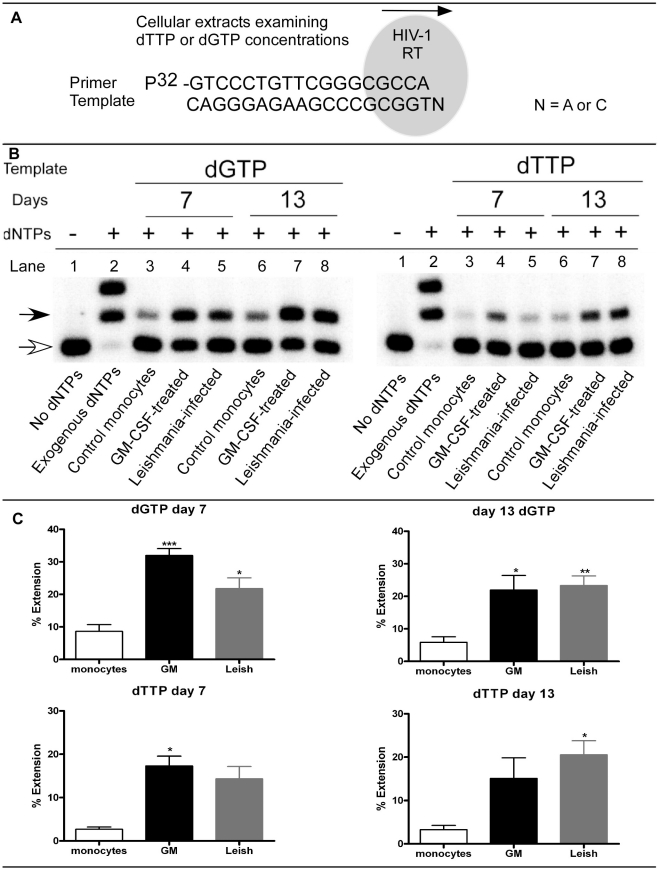
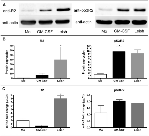
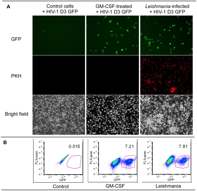
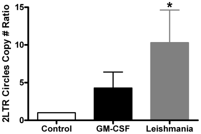
Leishmaniasis is a parasitic disease that is widely prevalent in many tropical and sub-tropical regions of the world. Infection with Leishmania has been recognized to induce a striking acceleration of Human Immunodeficiency Virus Type 1 (HIV-1) infection in coinfected individuals through as yet incompletely understood mechanisms. Cells of the monocyte/macrophage lineage are the predominant cell types coinfected by both pathogens. Monocytes and macrophages contain extremely low levels of deoxynucleoside triphosphates (dNTPs) due to their lack of cell cycling and S phase, where dNTP biosynthesis is specifically activated. Lentiviruses, such as HIV-1, are unique among retroviruses in their ability to replicate in these non-dividing cells due, at least in part, to their highly efficient reverse transcriptase (RT). Nonetheless, viral replication progresses more efficiently in the setting of higher intracellular dNTP concentrations related to enhanced enzyme kinetics of the viral RT. In the present study, in vitro infection of CD14+ peripheral blood-derived human monocytes with Leishmania major was found to induce differentiation, marked elevation of cellular p53R2 ribonucleotide reductase subunit and R2 subunit expression. The R2 subunit is restricted to the S phase of the cell cycle. Our dNTP assay demonstrated significant elevation of intracellular monocyte-derived macrophages (MDMs) dNTP concentrations in Leishmania-infected cell populations as compared to control cells. Infection of Leishmania-maturated MDMs with a pseudotyped GFP expressing HIV-1 resulted in increased numbers of GFP+ cells in the Leishmania-maturated MDMs as compared to control cells. Interestingly, a sub-population of Leishmania-maturated MDMs was found to have re-entered the cell cycle, as demonstrated by BrdU labeling. In conclusion, Leishmania infection of primary human monocytes promotes the induction of an S phase environment and elevated dNTP levels with notable elevation of HIV-1 expression in the setting of coinfection.


Conflict of interest statement

The authors have declared that no competing interests exist.

Figures

Figure 1
Figure 1. Determining cell viability.
A) 1×10⁶ monocytes/well were plated and then left untreated (control), GM-CSF-treated or Leishmania-infected at an MOI = 7. At various days afterwards, three wells/condition were collected, pooled and the cell numbers determined. Nine independent donors were examined. B–D) Images were captured using bright field microscope. Control cells had very few adherent cells as compared to GM-CSF-treated and Leishmania-infected cell cultures.
Figure 2
Figure 2. BrdU analysis.
A) Monocytes were infected with Leishmania at day 0. BrdU reagent was added 48 hours before fixing, processing and capturing the images. B) BrdU incorporation as monitored by FACS analysis. At day 13 of maturation, the cell populations from different groups were harvested and BrdU incorporation was determined. One of six independent donors is displayed for each group. C) The six independent donors were graphed and displayed as mean and SEM for BrdU+ cells. The GM-CSF-treated and Leishmania-infected MDMs have significantly higher BrdU incorporation as compared to control monocytes (* = p<0.05; ** = p<0.01).
Figure 3
Figure 3. HIV-1 RT based dNTP assay.
A) Diagram shows how a single nucleotide extension assay is done. The reaction contains the template, 5′ 32P-end-labeled primer, HIV-1 RT and cellular dNTP extract. After the reactions are completed, they are resolved on a polyacrylamide gel to determine product formation. The concentrations of dGTP and dTTP in the cellular extract will determine the amount of primer extension. B) For the different groups, primer extension products are shown for day 7 and day 13 cellular extracts. Control monocytes, GM-CSF-treated and Leishmania-infected cells are shown. The GM-CSF-treated cells are used as a positive control. Primer only (unextended) is indicated with an open arrow, whereas extended product formation is indicated with a filled arrow. The negative controls contain no dNTPs and are shown in lane 1. Positive controls (lanes 2) contained 50 µM of exogenous dNTP mix. Lanes 3–8 are cellular extracts from the different treatment groups. C) Graphs plotting the percent extension of dGTP and dTTP. From the primer extension assays, data was plotted for days 7 and 13 for dGTP (n = 9; top graphs) and for dTTP (n = 6; bottom graphs). Significantly different groups are displayed as * = p<0.05, ** = p<0.01 and *** = p<0.001 for the different groups as compared to control monocytes.
Figure 4
Figure 4. Western blot analysis of ribonucleotide reductase.
A) Cellular lysates from different treatment groups were analyzed for ribonucleotide reductase R2 and p53R2 subunits. Afterwards, blots were stripped and re-probed for actin. Freshly isolated monocytes (Mo), day 13 maturated GM-CSF (GM-CSF) and Leishmania (Leish) MDMs are shown. B) Quantitiation of western blots was done. Freshly isolated monocytes were set to 1 and increases in R2 and p53R2 expression levels for GM-CSF- and Leishmania-maturated MDMs groups are shown. Mean and SEM are displayed for four independent donors. C) qRT-PCR analysis was done on total cellular RNA extracts. mRNA fold changes for the different treatment groups (n = 3) are graphed as mean and SEM. Significantly different groups (p<0.05) as compared to monocyte control group are indicated with an asterisk (*).
Figure 5
Figure 5. HIV-1 D3 vector analysis.
A) Control, GM-CSF-treated and Leishmania-infected cells were transduced with HIV-1 D3 GFP vector. Twenty-four hours after transduction, the cells were examined for GFP expression (top row). Very few monocytes were GFP+, whereas more cells were GFP+ for the GM-CSF-treated and Leishmania-infected groups. For this experiment, the L. major were labeled with PKH dye and showed that the monocytes were infected. Bright field images were captured for the different groups. B) FACS analysis was done of three different cell populations. Data is representative of four different donors done at 24 h. Complete data sets for 24 h and 48 h are shown in Table 1.
Figure 6
Figure 6. 2LTR circle copy number ratio.
The different treatment groups were treated with the D3 GFP vector. At 48 h after transduction, total cellular DNA was collected and analyzed by real-time PCR. The control group for each donor was set to 1 and then compared to GM-CSF- and Leishmania-maturated MDMs groups. Mean and SEM are plotted. The significantly different group (p<0.05), as compared to control, is indicated with an asterisk. Seven independent donors were analyzed.

Similar articles

Cited by

References

    1. Alvar J, Aparicio P, Aseffa A, Den Boer M, Canavate C, et al. The relationship between leishmaniasis and AIDS: the second 10 years. Clin Microbiol Rev. 2008;21:334–359, table of contents. - PMC - PubMed
    1. Bern C, Maguire JH, Alvar J. Complexities of assessing the disease burden attributable to leishmaniasis. PLoS Negl Trop Dis. 2008;2:e313. - PMC - PubMed
    1. Alvar J, Yactayo S, Bern C. Leishmaniasis and poverty. Trends Parasitol. 2006;22:552–557. - PubMed
    1. Bates PA. Transmission of Leishmania metacyclic promastigotes by phlebotomine sand flies. Int J Parasitol. 2007;37:1097–1106. - PMC - PubMed
    1. Reithinger R, Brooker S, Kolaczinski JH. Visceral leishmaniasis in eastern Africa–current status. Trans R Soc Trop Med Hyg. 2007;101:1169–1170. - PMC - PubMed

Publication types

MeSH terms