. 2012;7(4):e34252.
doi: 10.1371/journal.pone.0034252. Epub 2012 Apr 4.

Epidermal transglutaminase (TGase 3) is required for proper hair development, but not the formation of the epidermal barrier

Affiliations

Epidermal transglutaminase (TGase 3) is required for proper hair development, but not the formation of the epidermal barrier

Susan John et al. PLoS One. 2012.

Abstract

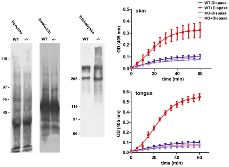
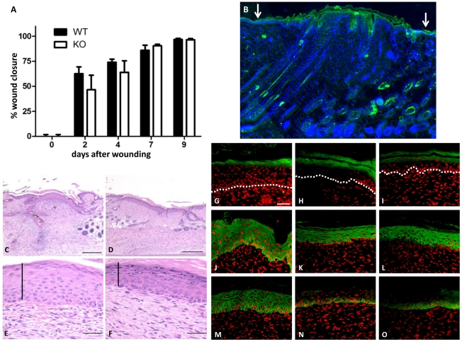
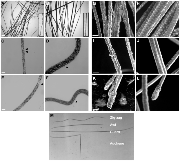
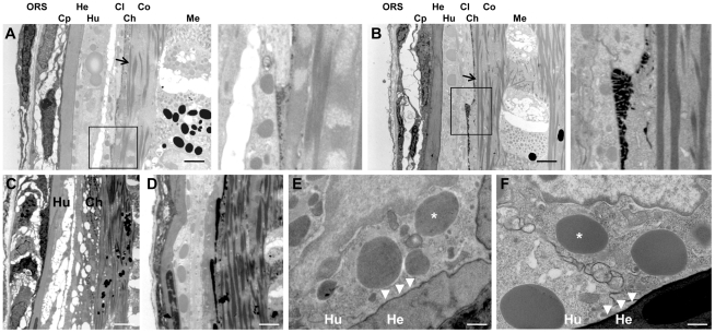
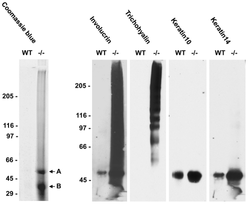
Transglutaminases (TGase), a family of cross-linking enzymes present in most cell types, are important in events as diverse as cell-signaling and matrix stabilization. Transglutaminase 1 is crucial in developing the epidermal barrier, however the skin also contains other family members, in particular TGase 3. This isoform is highly expressed in the cornified layer, where it is believed to stabilize the epidermis and its reduction is implicated in psoriasis. To understand the importance of TGase 3 in vivo we have generated and analyzed mice lacking this protein. Surprisingly, these animals display no obvious defect in skin development, no overt changes in barrier function or ability to heal wounds. In contrast, hair lacking TGase 3 is thinner, has major alterations in the cuticle cells and hair protein cross-linking is markedly decreased. Apparently, while TGase 3 is of unique functional importance in hair, in the epidermis loss of TGase 3 can be compensated for by other family members.


Conflict of interest statement

Competing Interests: The authors have declared that no competing interests exist.

Figures

Figure 1
Figure 1. Ablation of the TGM3 gene.
The locus of exons 4 to 7 of the TGM3 gene (A). The targeting construct was produced by insertion of the neomycin resistance cassette into exon 6. Southern blot analysis of cells after restriction digestion using NheI for the 5′ probe and PstI with the 3′ probe was used to identify correctly targeted ES cells. The disruption of exon 6 resulted in the wild type 28.8 kb NheI fragment being reduced to 3.4 kb, and the 5.3 kb PstI fragment being reduced to 4.9 kb. Southern blot analysis of ES cell DNA after NheI digestion and hybridization with the 5′ probe (B, left panel), and after PstI digestion and hybridization with the 3′ probe (B, right panel).
Figure 2
Figure 2. Gross phenotype of TGM3 null mice.
Southern blot analysis of tail biopsies from mice born of TGM3+/− interbreeding showed that homozygous mutants were born at the expected Mendelian ratios (A). The pelage hair of homozygous animals at 4 weeks of age showed a distinctive wavy pattern (B and C). This became less obvious as the mice matured (D). While gross hair abnormalities disappear with time, irregularities in the vibrissae, which are evident perinatally (E, mutant; F, control at P5) persist throughout life. Expression of TGase 3 is observed in pelage hair in the medulla and in the inner root sheath (G).
Figure 3
Figure 3. TGase 3 expression in epithelia.
Upper panel, protein extracts separated by SDS-PAGE were incubated with a mouse monoclonal antibody (A) and a rabbit polyclonal antiserum (B) against TGase 3. The expected TGase 3 band of 77 kDa was observed in extracts from wild type animals and was especially strong in oesophagus and tongue epithelium. In protein extracts from homozygous animals the signal was completely absent. The 50 kDa band seen in (A) corresponds to the heavy chain of IgG. Lower panel, immunofluorescence analysis of tongue (C, D) and back skin (E–L) of wild type (C, E, G, I, K) and TGM3 −/− animals (D, F, H, J, L). The sections were incubated with rabbit polyclonal antibodies against TGase 3 (C–F), TGase 6 (K and L) and monoclonal antibodies against TGase 1 (G, H) and TGase 2 (I and J). (scale bars represent 100 µm, the dotted line marks the dermal-epidermal junction).
Figure 4
Figure 4. Barrier function of the skin in TGM3−/− mice.
Transmission electron microscopy of skin from TGM3 −/− (A, C and E) and wild type (B, D and F) 4 week old animals. Cornified cell envelopes (closed arrowheads in A and B) and keratohyalin granules (*) were visible in mice of both genotypes and in some sections. The latter could be seen coalescing with the forming cornified envelope of granular layer keratinocytes (open arrowheads in C and D). The cells of the stratum corneum in both wild type and TGM3−/− animals consisted of a defined cell envelope surrounding compact, electron-dense cytoplasm containing condensed tonofibrils. Lucifer yellow failed to penetrate through the cornified envelope in either the newborn TGM3 −/− (G) or wild type skin (H). (scale bars B 0.5 µm, D 7 µm, F and H 50 µm). At birth there was no retention of toluidine blue dye in the skin of either TGM3−/− or wild type neonates (I). Dye was retained in the basal region of the whiskers in TGM3 −/− (L) neonates, a finding not seen in wild type mice (K). While formation of the epithelial barrier had occurred in control mice at E17.5, toluidine blue penetrated the skin of TGM3 −/− litter mates (J). Sonication of corneocytes isolated from skin biopsy punches for various times revealed that TGM3−/− corneocytes were more susceptible to lysis (M). The number shown is that of intact corneocytes remaining as a percentage of those initially isolated (n = 4).
Figure 5
Figure 5. Protein extractability and transglutaminase activity in TGM3−/− tissue.
Proteins from tissue extracts of TGM3 −/− and control skin were separated on an 8% SDS-PAGE and transferred to nitrocellulose. Probing with antibodies against involucrin revealed no alteration between the two genotypes, however the minor skin component trichohyalin showed an increased extractability (A). Protein extracts from wild type and TGM3 −/− skin and tongue were analyzed for biotin-cadaverin incorporation into NN′dimethylcasein, coated onto microtitre plates. Zymogen activation of the protein extract with dispase induced activity only in extracts from wild type mice (B).
Figure 6
Figure 6. Wound healing in TGM3−/− skin.
The rate of wound closure in TGM3 −/− mice was not different compared to control animals (A) (n = 9), despite an increase in TGase 3 expression in healing epidermis (B). At day 9 post wounding the thickness of the epidermal layer in the wounded region was greater in wild type skin (C, E) when compared to mutant skin (D, F). Staining of healing skin with keratinocyte differentiation markers in wild type (G, J, M) and TGM3−/− (H, I, K, L, N, O) animals demonstrated that while there was no change in the expression pattern of the keratinocyte markers loricrin (G–I) or keratin10 (J–L) between the mouse lines, keratin14 (M–O) already showed a more restricted staining pattern reminiscent of unwounded skin. (scale bars C, D 400 µm, E–G 100 µm)
Figure 7
Figure 7. Morphological changes in pelage hair lacking TGase 3.
Whole-mount light microscopy of TGM3 −/− (A) and wild type (B) hair (scale bar 10 µm). Inserts show higher magnification of lower regions of the hairs. Hair incubated overnight at 65°C with agitation in 2% SDS and 20 mM dithiothreitol (C–F) TGM3 −/− (C, E) and control hairs (D, F). Arrowheads indicate regions lacking or retaining the cuticle cells in the mutant and control hairs, respectively. Scanning electron microscopy of mutant (G, I) and control (H, J) hair shafts shows severe distortion of the hair, in particular the cuticle, in the absence of TGase 3. In contrast, the roots of the mutant hairs (K) appear similar to the wild type ones (L) (scale bar 20 µm). Each of the four main pelage hair types was present in the mutant animals (M).
Figure 8
Figure 8. Transmission electron microscopy of sections through the hair follicle of TGM3−/− (A, C) and wild type hair (B, D) (scale bar 10 µm).
Arrows show the keratin filaments. The cuticle layer is distorted in the mutant (magnified region), and disruption of the Huxley's layer is evident. Trichohyalin droplets (*) are seen as non-membrane-bound inclusions, in Huxley's layer of the IRS in both TGM3−/− (E) and control mice (F) and cornification occurs on the Henle's layer (arrow heads) (scale bar 2 µm). ORS-outer root sheath, Cp-companion layer, He-Henle's layer, Hu-Huxley's layer, Cl-cuticle of inner root sheath, Ch-hair cuticle, Co-hair cortex, Me-hair medulla.
Figure 9
Figure 9. Protein extractability in TGM3−/− hair.
Proteins were extracted from wild type and TGM3−/− hair with 2% SDS and 5% β-mercaptoethanol over night prior to SDS-PAGE separation and transferred to nitrocellulose. Membranes were probed with polyclonal antibodies against involucrin, trichohyalin, keratin10 and keratin14. Two prominent bands observed in the Coomassie stained SDS-PAGE of TGM3−/− protein lysates were identified by tryptic peptide mass fingerprinting as keratin6hf (A) and keratin17 (B).

Similar articles

Cited by

References

    1. Aeschlimann D, Thomazy V. Protein crosslinking in assembly and remodelling of extracellular matrices: the role of transglutaminases. Connect Tissue Res. 2000;41(1):1–27. - PubMed
    1. Aeschlimann D, Paulsson M. Transglutaminases: protein cross-linking enzymes in tissues and body fluids. Thromb Haemost. 1994;71(4):402–15. - PubMed
    1. Mycek MJ, Clarke DD, Neidle A, Waelsch H. Amine incorporation into insulin as catalyzed by transglutaminase. Arch Biochem Biophys. 1959;84:528–40. - PubMed
    1. Vader LW, de Ru A, van der Wal Y, Kooy YM, Benckhuijsen W, et al. Specificity of tissue transglutaminase explains cereal toxicity in celiac disease. J Exp Med. 2002;195(5):643–9. - PMC - PubMed
    1. Folk JE, Cole PW. Structural Requirements Of Specific Substrates For Guinea Pig Liver Transglutaminase. J Biol Chem. 1965;240:2951–60. - PubMed

Publication types

MeSH terms