. 2012;7(4):e34396.
doi: 10.1371/journal.pone.0034396. Epub 2012 Apr 4.

Propofol-induced changes in neurotrophic signaling in the developing nervous system in vivo

Affiliations

Propofol-induced changes in neurotrophic signaling in the developing nervous system in vivo

Jelena Popic et al. PLoS One. 2012.

Abstract

Several studies have revealed a role for neurotrophins in anesthesia-induced neurotoxicity in the developing brain. In this study we monitored the spatial and temporal expression of neurotrophic signaling molecules in the brain of 14-day-old (PND14) Wistar rats after the application of a single propofol dose (25 mg/kg i.p). The structures of interest were the cortex and thalamus as the primary areas of anesthetic actions. Changes of the protein levels of the brain-derived neurotrophic factor (BDNF) and nerve growth factor (NGF), their activated receptors tropomyosin-related kinase (TrkA and TrkB) and downstream kinases Akt and the extracellular signal regulated kinase (ERK) were assessed by Western immunoblot analysis at different time points during the first 24 h after the treatment, as well as the expression of cleaved caspase-3 fragment. Fluoro-Jade B staining was used to follow the appearance of degenerating neurons. The obtained results show that the treatment caused marked alterations in levels of the examined neurotrophins, their receptors and downstream effector kinases. However, these changes were not associated with increased neurodegeneration in either the cortex or the thalamus. These results indicate that in the brain of PND14 rats, the interaction between Akt/ERK signaling might be one of important part of endogenous defense mechanisms, which the developing brain utilizes to protect itself from potential anesthesia-induced damage. Elucidation of the underlying molecular mechanisms will improve our understanding of the age-dependent component of anesthesia-induced neurotoxicity.


Conflict of interest statement

Competing Interests: The authors have declared that no competing interests exist.

Figures

Figure 1
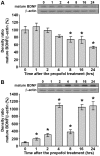
Figure 1. Propofol treatment differentially affects mature BDNF protein levels in PND14 rats.
Western blot analysis was used to determine the expression of mature BDNF in the cortex (A) and thalamus (B). Graphs that show changes in protein levels are accompanied by representative immunoblots. The data are expressed as percentages relative to the respective controls (mean ± SEM): *p<0.05 vs. control.
Figure 2
Figure 2. Propofol treatment differentially affects protein levels of total and phosphorylated TrkB in PND14 rats.
Western blot analysis was used to determine the expression of pTrkB and TrkB receptors in the cortex (A and B, respectively) and thalamus (C and D, respectively). Each graph is accompanied by a representative immunoblot. The data are expressed as percentages relative to the respective controls (mean ± SEM): *p<0.05 vs. control.
Figure 3
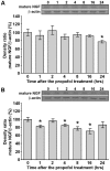
Figure 3. Propofol treatment down-regulates mature NGF protein levels in PND14 rats.
Western blot analysis was used to determine the expression of mature NGF in the cortex (A) and thalamus (B). Graphs that show changes in protein levels are accompanied by representative immunoblots. The data are expressed as percentages relative to the respective controls (mean ± SEM): *p<0.05 vs. control.
Figure 4
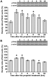
Figure 4. Propofol treatment up-regulates phosphorylated TrkA protein levels in PND14 rats.
Western blot analysis was used to determine the expression of pTrkA in the cortex (A) and thalamus (B). Graphs that show changes in protein levels are accompanied by representative immunoblots. The data are expressed as percentages relative to the respective controls (mean ± SEM): *p<0.05 vs. control.
Figure 5
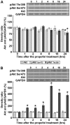
Figure 5. Propofol treatment differentially affects protein levels of total and phosphorylated Akt in PND14 rats.
Western blot analysis was used to determine the expression of Akt and pAkt (Thr308 and Ser473) kinase in the cortex (A) and thalamus (B). Each graph is accompanied by representative immunoblots. The data are expressed as percentages relative to the respective control (mean ± SEM): *p<0.05 vs. control.
Figure 6
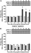
Figure 6. Propofol treatment differentially affects protein levels of total and phosphorylated ERK1/2 in PND14 rats.
Western blot analysis was used to determine the expression of ERK1/2 and pERK1/2 kinase in the cortex (A) and in the thalamus (B). Each graph is accompanied by representative immunoblots. The data are expressed as percentages relative to the respective control (mean ± SEM): *p<0.05 vs. control.
Figure 7
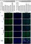
Figure 7. Propofol treatment did not induce neurodegeneration even with detected caspase-3 active fragment in PND14 rats.
Expression of the caspase-3 active fragment in the cortex (A) and thalamus (B). The data are expressed as percentages relative to the respective control (mean ± SEM), *p<0.05 vs. control. (C) Fluoro-Jade B staining (left panel), Hoechst 33258 staining (middle panel) and merged images (right panel) of representative brain sections of the cortex and the thalamus from control-(1st and 3rd row, respectively) and propofol-treated (16 h) (2nd and 4th row, respectively) PND14 animals. PND7 rats that served as a positive controls were treated with two i.p. doses (0.5 mg/kg) of (+) MK-801 and killed 24 h after the first administration of the drug. Degenerating neurons are marked with arrows and blood vessels with arrowheads. Scale bar = 20 µm.

Similar articles

Cited by

References

    1. Marik PE. Propofol: therapeutic indications and side-effects. Curr Pharm Des. 2004;10:3639–3649. - PubMed
    1. Krasowski MD, Harrison NL. General anaesthetic actions on ligand-gated ion channels. Cell Mol Life Sci. 1999;55:1278–1303. - PMC - PubMed
    1. Varju P, Katarova Z, Madarasz E, Szabo G. GABA signalling during development: new data and old questions. Cell Tissue Res. 2001;305:239–246. - PubMed
    1. Fredriksson A, Pontén E, Gordh T, Eriksson P. Neonatal exposure to a combination of N-methyl-D-aspartate and gamma-aminobutyric acid type A receptor anesthetic agents potentiates apoptotic neurodegeneration and persistent behavioral deficits. Anesthesiology. 2007;107:427–436. - PubMed
    1. Huang EJ, Reichardt LF. Neurotrophins: roles in neuronal development and function. Annu Rev Neurosci. 2001;24:677–736. - PMC - PubMed

Publication types

MeSH terms