. 2012;7(4):e34462.
doi: 10.1371/journal.pone.0034462. Epub 2012 Apr 5.

Circadian behaviour in neuroglobin deficient mice

Affiliations

Circadian behaviour in neuroglobin deficient mice

Christian A Hundahl et al. PLoS One. 2012.

Abstract

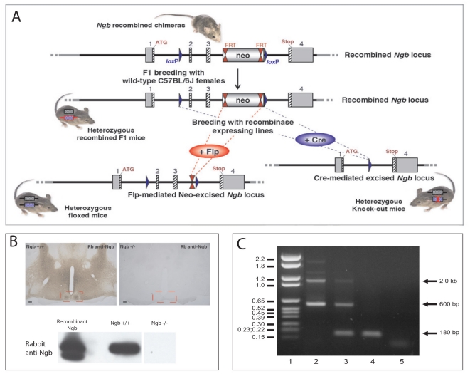
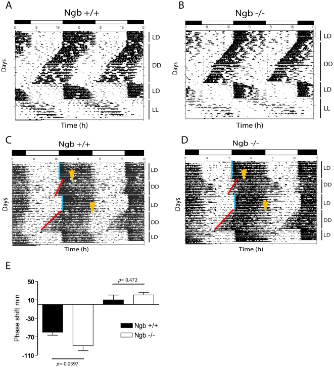
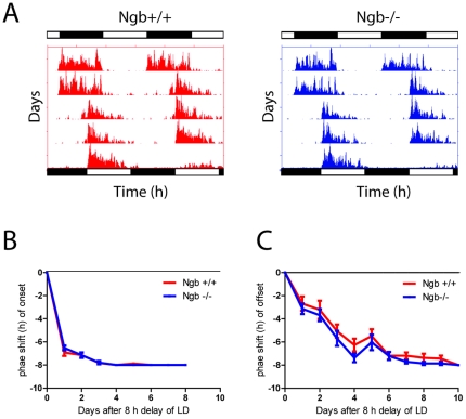
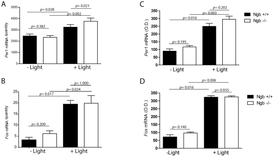
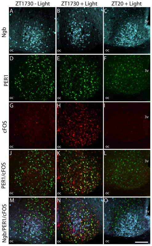
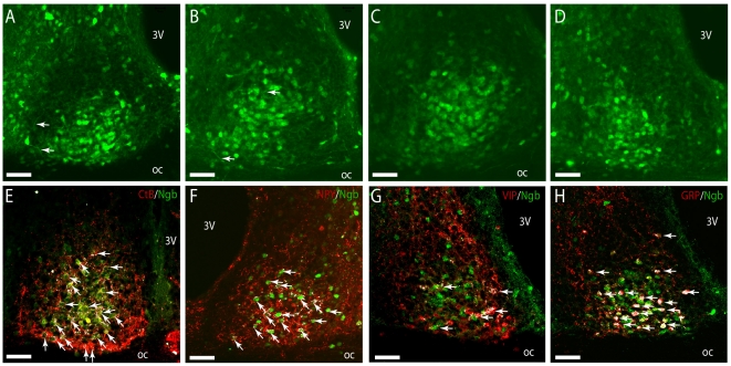
Neuroglobin (Ngb), a neuron-specific oxygen-binding globin with an unknown function, has been proposed to play a key role in neuronal survival. We have previously shown Ngb to be highly expressed in the rat suprachiasmatic nucleus (SCN). The present study addresses the effect of Ngb deficiency on circadian behavior. Ngb-deficient and wild-type (wt) mice were placed in running wheels and their activity rhythms, endogenous period and response to light stimuli were investigated. The effect of Ngb deficiency on the expression of Period1 (Per1) and the immediate early gene Fos was determined after light stimulation at night and the neurochemical phenotype of Ngb expressing neurons in wt mice was characterized. Loss of Ngb function had no effect on overall circadian entrainment, but resulted in a significantly larger phase delay of circadian rhythm upon light stimulation at early night. A light-induced increase in Per1, but not Fos, gene expression was observed in Ngb-deficient mice. Ngb expressing neurons which co-stored Gastrin Releasing Peptide (GRP) and were innervated from the eye and the geniculo-hypothalamic tract expressed FOS after light stimulation. No PER1 expression was observed in Ngb-positive neurons. The present study demonstrates for the first time that the genetic elimination of Ngb does not affect core clock function but evokes an increased behavioural response to light concomitant with increased Per1 gene expression in the SCN at early night.


Conflict of interest statement

Competing Interests: The authors have declared that no competing interests exist.

Figures

Figure 1
Figure 1. Generation and characterization of Ngb deficient mice.
A. Schematic representation of the targeting construct introduced into 129Sv/Pas embryonic stem (ES) cells to generate Ngb-knockout mice. Chimeric mice were crossed with Flp recombinase-expressing mice to remove the Neo resistance cassette and obtain F1 founder mice. F1 founder mice were crossed with mice expressing Cre recombinase ubiquitously which resulted in the deletion of Ngb exons 2 and 3 (B–C). B. Coronal sections of Ngb+/+ and Ngb−/− mice stained with rabbit anti-Ngb antiserum demonstrate specific staining in the Ngb wild type mouse and no staining in Ngb deficient mice. Similar results are obtained when tissue extracts from brains from the two genotypes were analyzed by western blotting (lower panel B). C represents PCR genotyping of littermates obtained by crossing heterozygous Ngb mice. Lane 1: DNA molecular marker VI (Roche), the sizes in kb are indicated into the left, lane 2: PCR on DNA from +/+ mouse, the expected size of bands are 2.0 kb and 600 bp, lane 3: PCR on DNA from +/− mouse, lane 4: PCR on DNA from Ngb −/− mouse, expected band size 180 bp; lane 5: Non-template control.
Figure 2
Figure 2. Running wheel activity in Ngb deficient mice.
A. Double plot actogram representing running wheel activity of Ngb wild type (A) and Ngb deficient mice (B) during a period of 12:12 h LD cycle followed by constant darkness (DD), re-entrainment to a new LD cycle followed by a period of constant light (LL). Both groups behave similarly and have a TAU not significantly different between the two genotypes. C and D. Light stimulation at ZT16 and ZT22 result in phase delays and phase advance, respectively which is illustrated in wild type animals (C) and in Ngb deficient mice (D). Ngb deficient mice display a significantly larger phase delay at ZT16 as shown in E. Yellow arrows indicates the time of the light pulse. Error bars = S.E.M., Mann Whitney-U test.
Figure 3
Figure 3. Re-entrainment after eight h phase shift of the LD cycle (jetlag) in Ngb deficient mice.
A. Representative actogram of Ngb wild type (red) and Ngb deficient mice (blue) entrained to a 12:12 h LD cycled followed by a eight h shift (delay) of the LD cycle. Bars in top of each actogram represent the LD cycle before the shift, the bars below the LD cycle after the shift. B. Quantitative analysis of the eight h phase delay of the LD cycle using the onset as phase marker (n = 7 of each genotype). Note that both groups re-entrain within three cycles. C. Quantitative analysis of the eight h phase delay of the LD cycle using the offset as phase marker (n = 7 of each genotype). Note both groups re-entrain within seven cycles.
Figure 4
Figure 4. Light induced expression of Per1 (A and C) and cFos (B and D) mRNA in Ngb deficient mice during early night.
Left panels (A–B) show the results of quantitative analysis using RT-PCR on SCN tissue ((mRNA/ß2MG mRNA (arbitrary units), see material and methods) and the results in the right panel (C and D) are obtained by semi-quantitative in situ hybridization (optical density, see material and methods). AB. A 30 min light pulse induced Per1 expression in Ngb deficient mice compared to wild type controls which is significant determined by RT-PCR on SCN tissue. Light stimulation significantly induces cFos expression in both genotypes but no difference was found in between the two genotypes (B and D). Error bars = S.E.M., Mann Whitney-U test.
Figure 5
Figure 5. Light induced cFOS and PER1 in Ngb expressing neurons of the mouse SCN.
The first column shows sections of mid SCN from a mouse euthanized at ZT 1730 without light stimulation. Ngb-IR (light blue) A, PER1 (green) D and cFOS (red) G are shown. Expression of Ngb-IR and PER1, but not cFOS-IR can been observed. Merged images J–M shows PER1/cFOS and PER1/cFOS/Ngb-IR, respectively. There was no co-expression between PER1 and Ngb. Similarly, in the mid column SCN from a mouse euthanized 90 min after light stimuli is shown. Note strong expression of cFOS (H) and a high degree of co-localization (yellow) with PER1-IR (K). No co-expression could be observed between Ngb and PER1-IR, but a subpopulation of Ngb-IR cells in the core was found to be cFOS positive (N). In the last column SCN from a mouse euthanized at ZT 20 after having received a 30 min light stimulus at ZT 16 is shown. Note cFOS-IR has disappeared (I) and substantially less PER1-IR (L) is expressed in both the nuclei and cytoplasm. No co-expression between Ngb-IR and PER1-IR could be seen (O). Oc; optic chiasm; 3 V, 3rd ventricle. Scale bar 100 µm.
Figure 6
Figure 6. Innervation and co-expression of Ngb in the mouse SCN.
In A–D Ngb-IR (green) can be seen in both the neuronal cell body and processes throughout the mid and ventral part of the SCN. Highest expression of Ngb-IR was observed in the mid/core part of SCN from rostral (A), mid (B–C) and in the ventral-lateral part in the caudal SCN (D). In (E) visual input from the RHT is depicted with cholera toxin subunit B (Ctb) (red). A high degree of innervation of Ngb-IR cells (green) was observed in the ventral and mid part of the SCN shown with white colour (arrows). Likewise Ngb-IR cells were also innervated by NPY-IR fibres (red) originating from the GHT (arrows) (F). In colchicine treated mice no co-expression of Ngb-IR and VIP-IR could be seen (G). Most GRP-IR cells were seen to co-express Ngb-IR (arrows) in colchicine treated mice (H). Oc; optic chiasm; 3 V, 3rd ventricle. Scale bars 50 µm.

References

    1. Golombek DA, Rosenstein RE. Physiology of circadian entrainment. Physiol Rev. 2010;90:1063–1102. - PubMed
    1. Antle MC, Smith VM, Sterniczuk R, Yamakawa GR, Rakai BD. Physiological responses of the circadian clock to acute light exposure at night. Rev Endocr Metab Disord. 2009;10:279–291. - PubMed
    1. Reppert SM, Weaver DR. Molecular analysis of mammalian circadian rhythms. Annu Rev Physiol. 2001;63:647–676. - PubMed
    1. Yan L, Silver R. Resetting the brain clock: time course and localization of mPER1 and mPER2 protein expression in suprachiasmatic nuclei during phase shifts. Eur J Neurosci. 2004;19:1105–1109. - PMC - PubMed
    1. Hannibal J, Fahrenkrug J. Neuronal input pathways to the brain's biological clock and their functional significance. Adv Anat Embryol Cell Biol. 2006;182:1–71. - PubMed

Publication types

MeSH terms

LinkOut - more resources