. 2012;7(4):e34514.
doi: 10.1371/journal.pone.0034514. Epub 2012 Apr 4.

F-actin-dependent regulation of NESH dynamics in rat hippocampal neurons

Affiliations

F-actin-dependent regulation of NESH dynamics in rat hippocampal neurons

Jeomil Bae et al. PLoS One. 2012.

Abstract

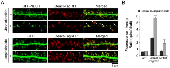
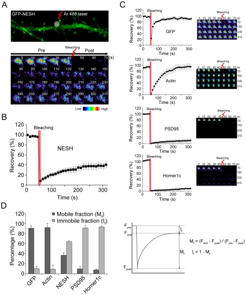
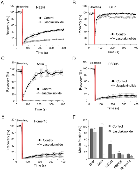
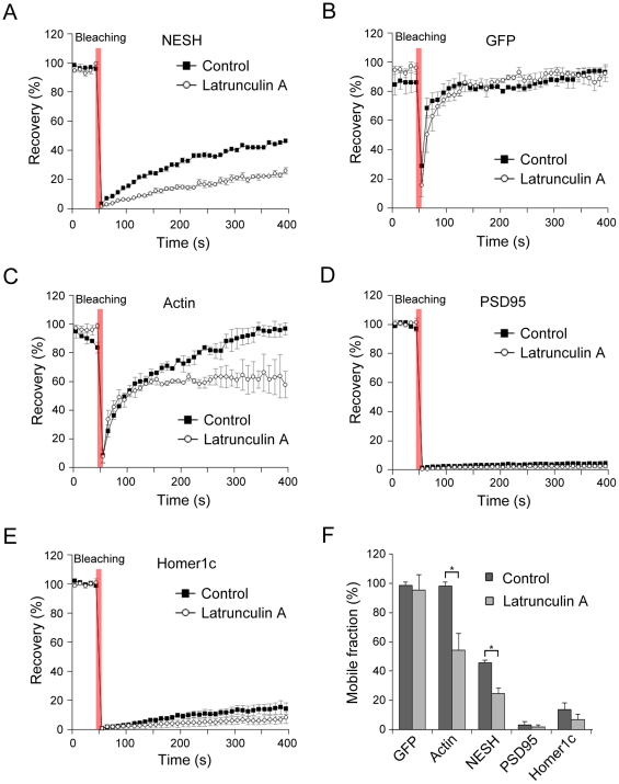
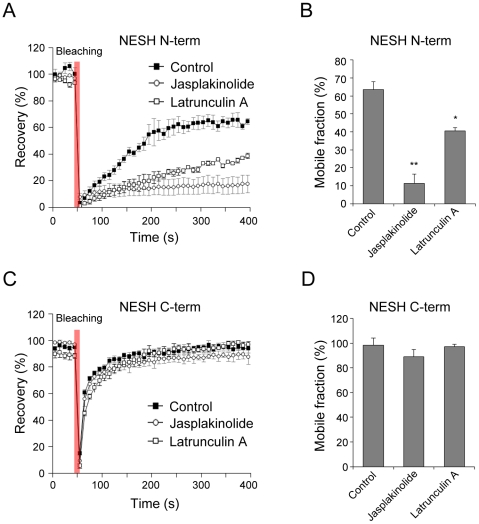
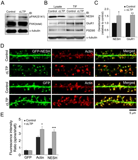
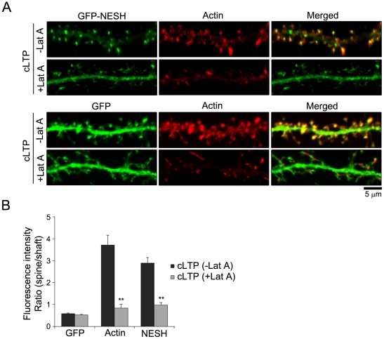
Synaptic plasticity is an important feature of neurons essential for learning and memory. Postsynaptic organization and composition are dynamically remodeled in response to diverse synaptic inputs during synaptic plasticity. During this process, the dynamics and localization of postsynaptic proteins are also precisely regulated. NESH/Abi-3 is a member of the Abl interactor (Abi) protein family. Overexpression of NESH is associated with reduced cell motility and tumor metastasis. Strong evidence of a close relationship between NESH and the actin cytoskeleton has been documented. Although earlier studies have shown that NESH is prominently expressed in the brain, its function and characteristics are yet to be established. Data from the present investigation suggest that synaptic localization of NESH in hippocampal neurons is regulated in an F-actin-dependent manner. The dynamic fraction of NESH in the dendritic spine was analyzed using FRAP (fluorescence recovery after photobleaching). Interestingly, F-actin stabilization and disruption significantly affected the mobile fraction of NESH, possibly through altered interactions of NESH with the F-actin. In addition, NESH was synaptically targeted from the dendritic shaft to spine after induction of chemical LTP (long-term potentiation) and the translocation was dependent on F-actin. Our data collectively support the significance of the F-actin cytoskeleton in synaptic targeting of NESH as well as its dynamics.


Conflict of interest statement

Competing Interests: The authors have declared that no competing interests exist.

Figures

Figure 1
Figure 1. F-actin-dependent synaptic translocation of NESH in hippocampal neurons.
(A) The effect of F-actin stabilization on localization of NESH was investigated. Hippocampal neurons were co-transfected with GFP-NESH (or GFP) and pLifeact-TagRFP at 10–12 DIV. pLifeact-TagRFP was employed to image the F-actin cytoskeleton within cells. Transfected neurons at 16–18 DIV were treated with jasplakinolide (5 µM for 10 min), fixed, and imaged. Colocalization between NESH and F-actin is indicated with white arrows in the merged image. (B) Quantitative analysis of the intensity ratio of spines vs. shafts from data obtained in Fig. 1A (N = 16 neurons for each condition). Data are presented as means ± SEM. ***p<0.001.
Figure 2
Figure 2. FRAP analysis of NESH dynamics in dendritic spines.
(A) To investigate the dynamics of NESH in a single spine, the FRAP (fluorescence recovery after photobleaching) assay was performed. Hippocampal neurons at 10–12 DIV were transfected with GFP-NESH and subjected to FRAP imaging at 16–18 DIV. A single spine of GFP-NESH-transfected neurons was set to ROI (region of interest) and bleached for 7 s with an Ar 488 laser, and recovery of GFP-NESH observed at intervals of 10 s over a time-course of 5 min. (B) Recovery curve of GFP-NESH from data obtained in Fig. 2A. NESH fluorescence was slowly recovered (up to ∼40%) for 5 min (N = 15, data from three to five independent experiments). (C) Hippocampal neurons at 10–12 DIV were transfected with GFP, GFP-actin, GFP-PSD95 or GFP-Homer1c, and used for FRAP imaging at 16–18 DIV. NESH mobility was compared with that of other proteins using FRAP (N = 15 for each protein, data from three to five independent experiments). (D) Analysis of mobile/immobile fractions from data obtained in Fig. 2B and C. Fend: fluorescence at the end time-point, Fpost: fluorescence right after photobleaching, Fpre: fluorescence before photobleaching, Mf: mobile fraction, If: immobile fraction. Data are presented as means ± SEM.
Figure 3
Figure 3. Effect of F-actin stabilization on NESH mobility in dendritic spines.
FRAP analysis was performed to examine the effect of F-actin stabilization on NESH mobility. Hippocampal neurons at 10–12 DIV were transfected with GFP-NESH, GFP, GFP-actin, GFP-PSD95 or GFP-Homer1c, and subjected to FRAP analysis at 16–18 DIV. (A, F) Recovery curves of GFP-NESH in non-treated (control) and jasplakinolide-treated neurons (mobile fractions: 43.9±2.4% at 400 s for control, 13.6±2.8% at 400 s for jasplakinolide). (B, F) Recovery curves of GFP (mobile fractions: 92.7±8.0% at 400 s for control, 83.3±1.3% at 400 s for jasplakinolide). (C, F) Recovery curves of GFP-actin (mobile fractions: 98.6±8.7% at 400 s for control, 0.2±0.1% at 400 s for jasplakinolide). (D–F) Recovery curves and mobile fractions of the scaffolding proteins, PSD95 and Homer1c (control: 16.7±5.2% at 400 s for GFP-PSD95, 14.8±6.6% at 400 s for GFP-Homer1c, jasplakinolide: 12.0±2.7% at 400 s for GFP-PSD95, 7.0±4.2% at 400 s for GFP-Homer1c). (F) Analysis of the mobile/immobile fractions from data obtained in Fig. 3A–E (N = 15 for each condition, data from three to five independent experiments). Data are presented as means ± SEM. **p<0.01.
Figure 4
Figure 4. Effect of F-actin disruption on mobility of NESH.
To examine the effect of F-actin disruption on NESH mobility in the dendritic spine, FRAP analyses were performed using hippocampal neurons at 16–18 DIV that were transfected with GFP-NESH, GFP, GFP-actin, GFP-PSD95 and GFP-Homer1c at 10–12 DIV. F-actin disruption was induced by treating with latrunculin A (5 µM for 10 min). (A, F) Recovery curves of GFP-NESH in non-treated (control) and latrunculin A-treated neurons (mobile fractions: 45.6±1.9% at 400 s for control, 24.9±3.6% at 400 s for latrunculin A), suggesting that dynamic actin remodeling is crucial for the mobility and dynamics of NESH. (B, F) Recovery curves of GFP (mobile fractions: 98.2±2.2% at 400 s for control, 95.4±10.3% at 400 s for latrunculin A) (C, F) Recovery curves of GFP-actin (mobile fractions: 98.1±2.6% at 400 s for control, 54.2±11.4% at 400 s for latrunculin A) (D–F) Recovery curves and mobile fractions of scaffold proteins, GFP-PSD95 and GFP-Homer1c, showed no significant differences. (F) Analysis of mobile/immobile fractions from data obtained in Fig. 4A–E (N = 15 for each condition, data from three to five independent experiments). Data are presented as means ± SEM. *p<0.05.
Figure 5
Figure 5. Importance of the F-actin-binding region in NESH dynamics.
The dynamics of NESH-N-term (N-terminal half of NESH, F-actin binding region) and C-term (C-terminal half of NESH, F-actin non-binding region) were analyzed using FRAP. Hippocampal neurons at 10–12 DIV were transfected with GFP-NESH-N-term or GFP-NESH-C-term, and subjected to FRAP analysis at 16–18 DIV. (A, B) Recovery curves and mobile fractions of NESH-N-term (63.3±4.5% at 400 s for control, 11.2±5.1% for jasplakinolide, 40.4±1.7% for latrunculin A). N = 15 for each condition, data from three to five independent experiments. (C, D) Recovery curves and mobile fractions of NESH-C-term (98.1±6.0% for control, 89.0±5.7% at 400 s for jasplakinolide, 97.0±2.0% at 400 s for latrunculin A). N = 15 for each condition, data from three to five independent experiments. Data are presented as means ± SEM. *p<0.05, **p<0.01.
Figure 6
Figure 6. Synaptic translocation of NESH via LTP induction.
The glycine-induced chemical LTP (long-term potentiation) method was used to mimic physiological LTP. (A) Chemical LTP (cLTP) was induced in hippocampal neurons at 16–18 DIV, and the level of phospho-PAK assessed to ascertain cLTP induction. (B) After cLTP induction, the Triton X-100-insoluble fraction was extracted from hippocampal neurons, and the association between NESH and cytoskeleton or synaptic site examined using immunoblot analysis. (C) The band intensities were quantified and normalized by loading control (α-tubulin). (D) Synaptic translocation of NESH was examined during LTP. Hippocampal neurons at 10–12 DIV were transfected with GFP-NESH (or GFP). After cLTP induction at 16–18 DIV, transfected neurons were fixed and stained with Alexa 594-conjugated phalloidin, and NESH localization examined. GFP served as the control. (E) Analysis of the fluorescence intensity ratio in dendritic spine vs. shaft from data obtained in Fig. 6D (N = 12–16 neurons for each condition). Data are presented as means ± SEM. *p<0.05, **p<0.01, ***p<0.001.
Figure 7
Figure 7. F-actin-dependent synaptic translocation of NESH after LTP induction.
(A) Hippocampal neurons at 10–12 DIV were transfected with GFP-NESH (or GFP). To determine the importance of the F-actin cytoskeleton in NESH translocation during cLTP, transfected neurons at 16–18 DIV were treated with latrunculin A (5 µM for 10 min), and cLTP was subsequently induced. Following fixing of neurons, NESH localization was examined. (B) Analysis of the intensity ratio (spine vs. shaft) from data obtained in Fig. 7A (N = 11–13 neurons for each condition). Data are presented as means ± SEM. **p<0.01.

References

    1. Bourne JN, Harris KM. Balancing structure and function at hippocampal dendritic spines. Annual review of neuroscience. 2008;31:47–67. - PMC - PubMed
    1. Sheng M, Kim MJ. Postsynaptic signaling and plasticity mechanisms. Science. 2002;298:776–780. - PubMed
    1. McGee AW, Bredt DS. Assembly and plasticity of the glutamatergic postsynaptic specialization. Current opinion in neurobiology. 2003;13:111–118. - PubMed
    1. Sheng M, Hoogenraad CC. The postsynaptic architecture of excitatory synapses: a more quantitative view. Annual review of biochemistry. 2007;76:823–847. - PubMed
    1. Renner M, Specht CG, Triller A. Molecular dynamics of postsynaptic receptors and scaffold proteins. Current opinion in neurobiology. 2008;18:532–540. - PubMed

Publication types

MeSH terms