. 2012;7(4):e34672.
doi: 10.1371/journal.pone.0034672. Epub 2012 Apr 4.

GPR30, the non-classical membrane G protein related estrogen receptor, is overexpressed in human seminoma and promotes seminoma cell proliferation

Affiliations

GPR30, the non-classical membrane G protein related estrogen receptor, is overexpressed in human seminoma and promotes seminoma cell proliferation

Nicolas Chevalier et al. PLoS One. 2012.

Abstract

Background: Testicular germ cell tumours are the most frequent cancer of young men with an increasing incidence all over the world. Pathogenesis and reasons of this increase remain unknown but epidemiological and clinical data have suggested that fetal exposure to environmental endocrine disruptors (EEDs) with estrogenic effects, could participate to testicular germ cell carcinogenesis. However, these EEDs (like bisphenol A) are often weak ligands for classical nuclear estrogen receptors. Several research groups recently showed that the non classical membrane G-protein coupled estrogen receptor (GPER/GPR30) mediates the effects of estrogens and several xenoestrogens through rapid non genomic activation of signal transduction pathways in various human estrogen dependent cancer cells (breast, ovary, endometrium). The aim of this study was to demonstrate that GPER was overexpressed in testicular tumours and was able to trigger JKT-1 seminoma cell proliferation.

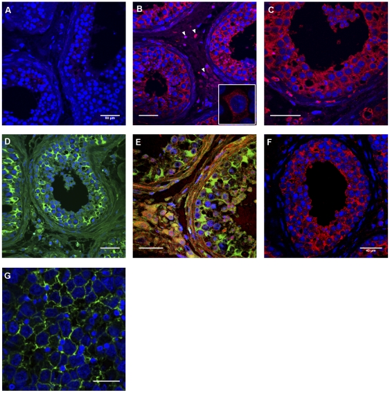
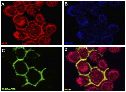
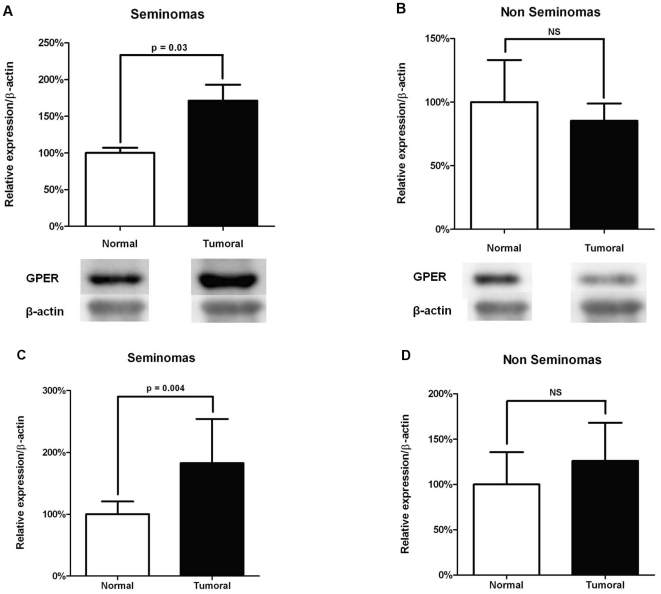
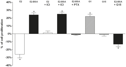
Results: We report here for the first time a complete morphological and functional characterization of GPER in normal and malignant human testicular germ cells. In normal adult human testes, GPER was expressed by somatic (Sertoli cells) and germ cells (spermatogonia and spermatocytes). GPER was exclusively overexpressed in seminomas, the most frequent testicular germ cell cancer, localized at the cell membrane and triggered a proliferative effect on JKT-1 cells in vitro, which was completely abolished by G15 (a GPER selective antagonist) and by siRNA invalidation.

Conclusion: These results demonstrate that GPER is expressed by human normal adult testicular germ cells, specifically overexpressed in seminoma tumours and able to trigger seminoma cell proliferation in vitro. It should therefore be considered rather than classical ERs when xeno-estrogens or other endocrine disruptors are assessed in testicular germ cell cancers. It may also represent a prognosis marker and/or a therapeutic target for seminomas.


Conflict of interest statement

Competing Interests: The authors have declared that no competing interests exist.

Figures

Figure 1
Figure 1. Immunofluorescence analysis of GPER expression in normal human testis and seminoma.
GPER was expressed (red fluorescence) in normal testis both in the interstitial compartment (white arrows) and in seminiferous tubules (B, C) as opposed to negative control (A). In order to specifically identify Sertoli cells in seminiferous tubules, a mouse monoclonal anti-vimentin clone V9 Ab (green fluorescence; Dako® Denmark A/S, Glostrup, Denmark) was used (D). Double staining (E) allowed identifying GPER expression both in germinal and Sertoli cells (yellow fluorescence [merge]). GPER was also expressed (red fluorescence) in seminoma cells (F), which were identified on a tumoural section (G) by using a mouse monoclonal antibody against placental alkaline phosphatase (green fluorescence; Thermo Fisher Scientific®, Fremont, CA, USA). In all sections, cell nuclei were stained by Hoechst 33258. Human MCF-7 breast cancer cells have been shown as positive control (window in B). Magnification: ×40 (A, B, D, E, F); ×63 (C, G).
Figure 2
Figure 2. Immunolocalization of GPER in JKT-1 seminoma cells.
In JKT-1 cells, GPER (red fluorescence) had both a membranous and intracytoplasmic perinuclear localization (A), whereas the classical oestrogen receptor ERβ had an intracytoplasmic perinuclear and nuclear localization (B, blue fluorescence). E2-BSA, an impermeable E2 conjugate that does not cross the membrane, stained the cell membrane when coupled to FITC (C, green fluorescence). By double staining (D), E2-BSA (green) and GPER (red) co-localized at the membrane (yellow [merge]); GPER was also expressed in the cytoplasm (red). (A, B, C, D: magnification, ×63).
Figure 3
Figure 3. GPER expression in seminoma and non seminoma tumours.
Quantitative analysis of GPER expression was performed by Western blotting (A, B) and RT-PCR (C, D) in testicular germ cell tumours. β-actin was taken in each case as a house-keeping gene both in Western blotting and RT-PCR. Relative expression of GPER mRNA and protein of the tumoural tissue was expressed as percentage of the value obtained in the normal peri-tumoural testicular tissue of the same testis (represented as 100%). Histograms show the mean and standard deviation of the mRNA and protein values for a group of 8 seminomas (A, C) and for a group of 7 non seminomas (B, D). Both mRNAs and protein values were significantly higher in seminomas when compared to normal peri-tumoural testicular tissue (A, C). At the opposite, mRNAs and protein values were not different (P>0.05) in non seminoma tumours when compared to normal peri-tumoural testicular tissue (B, D).
Figure 4
Figure 4. GPER triggers JKT-1 cell proliferation in vitro.
JKT-1 cells were seeded in six-well plates (0,6×10⁶ cells/well). After 48 h, the JKT-1 cells were washed and oestrogen starved overnight in phenol red-free DMEM supplemented with 1% charcoal-stripped FBS. Serum-deprived JKT-1 cells were then incubated for 24 h with E2-BSA (1 nM), an impermeable E2-conjugate, which cannot thus interact with classical nuclear or cytoplasmic estrogen receptors, after a pre-treatment with G15 (1 nM), a GPER antagonist, or ICI-182,780 (1 µM), a pure ER antagonist, or pertussis toxin (100 ng/mL), a GPCR protein inhibitor. G1 (1 nM), a GPER-specific agonist, was used as a positive control. E2 at the same concentration (1 nM), which induces an inhibitory effect on cell proliferation neutralized by ER antagonist ICI (1 µM), was used for comparison. Histograms represent percentages of variation in the JKT-1 cell number compared with the control (0%), which was measured at 1,2×10⁶ cells/well after 24 hours of incubation in the steroid-free medium with ethanol (10% of variation represent an increase or a decrease of 60 000 cells). All results are expressed as means ± SEM of three independents experiments. * P<0.01.
Figure 5
Figure 5. Effects of GPER knockdown on E2 and E2-BSA-induced JKT-1 cell proliferation.
(A) JKT-1 cells were transfected with 50 nM of siRNA designed to knockdown GPER or with control siRNA. After 72-h incubation, proteins were extracted and subjected to Western blotting to confirm the specific inhibitory activity of GPER siRNA after normalization with β-actin, which was evaluated as a house-keeping gene. (B) After 72 h transfection with 50 nM of GPER siRNA or control siRNA, JKT-1 cells were incubated for 24 h with E2 (1 nM) or E2-BSA (1 nM). Histograms represent the percentages of variation of the JKT-1 cell number compared to control without estrogens. Cell number was measured at 1,5×10⁶ cells/well after 24 hours of incubation in the steroid-free medium with ethanol (10% of variation representing an increase or a decrease of 90 000 cells). All results are expressed as means ± SEM of three independents experiments (*P<0.01).

References

    1. Skakkebaek NE, Rajpert-De Meyts E, Jorgensen N, Carlsen E, Petersen PM, et al. Germ cell cancer and disorders of spermatogenesis: an environmental connection? APMIS. 1998;106:3–11; discussion 12. - PubMed
    1. Carreau S, Bourguiba S, Lambard S, Galeraud-Denis I, Genissel C, et al. Reproductive system: aromatase and estrogens. Mol Cell Endocrinol. 2002;193:137–143. - PubMed
    1. Jones ME, Simpson ER. Oestrogens in male reproduction. Baillieres Best Pract Res Clin Endocrinol Metab. 2000;14:505–516. - PubMed
    1. Hardell L, Bavel B, Lindstrom G, Eriksson M, Carlberg M. In utero exposure to persistent organic pollutants in relation to testicular cancer risk. Int J Androl. 2006;29:228–234. - PubMed
    1. Kinugawa K, Hyodo F, Matsuki T, Jo Y, Furukawa Y, et al. Establishment and characterization of a new human testicular seminoma cell line, JKT-1. Int J Urol. 1998;5:282–287. - PubMed