. 2012;7(4):e34863.
doi: 10.1371/journal.pone.0034863. Epub 2012 Apr 9.

In vivo analysis of MEF2 transcription factors in synapse regulation and neuronal survival

Affiliations

In vivo analysis of MEF2 transcription factors in synapse regulation and neuronal survival

M Waseem Akhtar et al. PLoS One. 2012.

Abstract

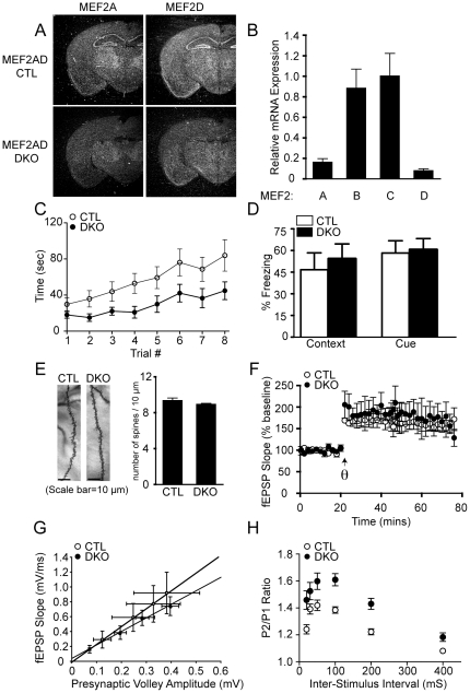
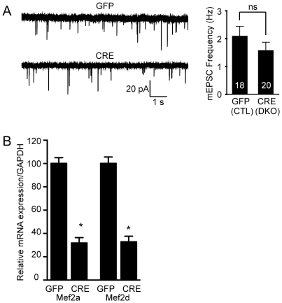
MEF2 (A-D) transcription factors govern development, differentiation and maintenance of various cell types including neurons. The role of MEF2 isoforms in the brain has been studied using in vitro manipulations with only MEF2C examined in vivo. In order to understand specific as well as redundant roles of the MEF2 isoforms, we generated brain-specific deletion of MEF2A and found that Mef2aKO mice show normal behavior in a range of paradigms including learning and memory. We next generated Mef2a and Mef2d brain-specific double KO (Mef2a/dDKO) mice and observed deficits in motor coordination and enhanced hippocampal short-term synaptic plasticity, however there were no alterations in learning and memory, Schaffer collateral pathway long-term potentiation, or the number of dendritic spines. Since previous work has established a critical role for MEF2C in hippocampal plasticity, we generated a Mef2a, Mef2c and Mef2d brain-specific triple KO (Mef2a/c/dTKO). Mef2a/c/d TKO mice have early postnatal lethality with increased neuronal apoptosis, indicative of a redundant role for the MEF2 factors in neuronal survival. We examined synaptic plasticity in the intact neurons in the Mef2a/c/d TKO mice and found significant impairments in short-term synaptic plasticity suggesting that MEF2C is the major isoform involved in hippocampal synaptic function. Collectively, these data highlight the key in vivo role of MEF2C isoform in the brain and suggest that MEF2A and MEF2D have only subtle roles in regulating hippocampal synaptic function.


Conflict of interest statement

Competing Interests: The authors have declared that no competing interests exist.

Figures

Figure 1
Figure 1. Strategy to generate a conditional MEF2A allele.
LoxP sites were inserted into introns 1 and 2 through homologous recombination. Cre-mediated excision results in one loxP site in the place of exon 2.
Figure 2
Figure 2. Mef2aKO mice show normal behavior.
(A) Left, PCR genotyping to distinguish different Mef2a alleles. Right, the bar graph shows expression levels of Mef2 isoforms in the hippocampus of 4 week old animals (n=3/genotype), detected by quantitative PCR. The fold change in RNA was calculated using the comparative Ct method, normalizing to GAPDH as a control. (B) Nissl staining of brain cross-sections from littermate control (CTL) and Mef2a KO mice at 2 months of age. Brain-specific deletion of MEF2A was achieved by crossing MEF2AloxP/loxP mice with mice harboring a transgene for hGFAP-Cre. (C) Mef2aKO (KO) mice show no significant differences in locomotor activity compared to littermate CTL mice as assessed by consecutive beam breaks over a two-hour period, although the number of beam breaks significantly decreased over time (F23,184=41.84, P<0.0001), there was no significant interaction between group and time (CTL, n=8; Mef2aKO mice, n=9). (D) Mef2aKO mice have no difference in motor coordination as assessed in the rotarod test compared to littermate CTL mice (CTL, n=8; Mef2aKO mice, n=9). (E) Mef2aKO mice exhibit normal anxiety-like behavior, as assessed in the open field test (CTL, n=8; Mef2aKO mice, n=9). (F) Context- and cue-dependent fear conditioning is unaltered in Mef2aKO mice relative to littermate CTL mice (CTL, n=8; Mef2aKO mice, n=9). All data is shown as mean ± SEM.
Figure 3
Figure 3. Brain-specific deletion of Mef2a and Mef2d causes impairments in motor coordination, as well as presynaptic release probability.
(A) Detection of Mef2a and Mef2d transcripts by in situ hybridization in littermate CTL and Mef2a/d DKO (DKO) mice. Arrow indicates hippocampus. (B) Expression level of MEF2 transcription factors in the hippocampus of Mef2a/d DKO mice as detected by quantitative RT-PCR. RNA was isolated from the hippocampus of 4 week old animals (n=3/genotype), and then expression determined by quantitative PCR. The fold change in RNA was calculated using the comparative Ct method, normalizing to GAPDH as a control. (C) Mef2a/d DKO mice exhibit impaired motor coordination as assessed by falling off the accelerating rotarod faster than littermate CTLs (F1,7=27.64, P<0.05; CTL, n=8; Mef2a/d DKO, n=10), posthoc analysis demonstrated that trial numbers 3–8 were significantly different between the Mef2a/d DKO and CTL mice. (D) Mef2a/d DKO mice show normal associative learning, as assessed by context- and cue-dependent fear conditioning (CTL, n=8; Mef2a/d DKO, n=10). (E) Representative images of Golgi-stained CA1 pyramidal neurons and a bar graph comparing the number of dendritic spines per 10 µm length of dendrite from littermate CTL and Mef2a/d DKO mice (n=31 dendritic segments from 31 neurons of two CTL mice and n=68 dendritic segments from 68 neurons of four Mef2a/d DKO mice). (F) CA1 LTP induction and maintenance is normal in DKO mice (CTL, n=3; Mef2a/d DKO, n=3). (G) Input-output relations are normal in Mef2a/d DKO mice (CTL, n=4; Mef2a/d DKO, n=4; solid lines indicate lines of best fit.). (H) Paired-pulse facilitation is increased in Mef2a/d DKO mice at Inter-Stimulus Interval 20, 30, 50, 100, 200 and 400 (mS) compared to littermate CTLs, suggesting decreased presynaptic release probability (CTL, n=8; Mef2a/d DKO, n=7). All data is shown as mean ± SEM.
Figure 4
Figure 4. The loss of MEF2A/D does not alter basal synaptic transmission.
(A) mEPSC frequency is unchanged upon deletion of Mef2a/d in hippocampal culture neurons using lentivirus expressing Cre recombinase (p>0.05). The number of recordings is shown in the bar graph for the GFP and Cre infected neurons. Data is shown as mean ± SEM. (B) Lentivirus containing either GFP or Cre was infected at 4 DIV in hippocampal culture neurons and cells were harvested at 15–18 DIV, in parallel with the time of recordings. Quantitative PCR was used to confirm the deletion of Mef2a/d (*p<0.05). Relative mRNA expression of Mef2a/d was normalized to GAPDH (GFP, n=6; Cre, n=6). Data is shown as mean ± SEM.
Figure 5
Figure 5. Brain-specific triple deletion of mef2a, c and d impairs survival, brain size and synaptic plasticity.
(A) Kaplan-Meier survival curves from littermate CTL and Mef2a/c/d TKO (TKO) mice. (B) Hematoxylin and eosin staining of coronal section from littermate CTLs and Mef2a/c/d TKO. (C) Representative TUNEL staining of sections from littermate CTL and Mef2a/c/d TKO brains at 1 month of age. The white arrows indicate TUNEL stained cells. Bar graph shows an increase in apoptotic cells in Mef2a/c/d TKO mice. Data is shown as mean ± SEM. (D) Representative images of Golgi-stained CA1 pyramidal neurons from CTL and Mef2a/c/d TKO (TKO) mice (Data pooled from 15 sections/2 CTL mice and from 38 sections/4 Mef2a/c/d TKO mice). (E) CA1 LTP induction and maintenance is normal in Mef2a/c/d TKO mice (CTL, n=5; Mef2a/c/d TKO, n=6). (F) The slope of the input-output functions was significantly lower in Mef2a/c/d TKO mice compared to littermates controls (P<0.05; CTL, n=10; Mef2a/c/d TKO, n=7; solid lines indicate lines of best fit). (G) Paired-pulse facilitation is significantly increased in Mef2a/c/d TKO mice at Inter-Stimulus Interval 50, 100, 200 and 400 (mS) compared to littermate CTLs (P<0.05; CTL, n=8; Mef2a/c/d TKO, n=7).

References

    1. Potthoff MJ, Olson EN. MEF2: a central regulator of diverse developmental programs. Development. 2007;134:4131–4140. - PubMed
    1. Lyons GE, Micales BK, Schwarz J, Martin JF, Olson EN. Expression of mef2 genes in the mouse central nervous system suggests a role in neuronal maturation. J Neurosci. 1995;15:5727–5738. - PMC - PubMed
    1. Mao Z, Bonni A, Xia F, Nadal-Vicens M, Greenberg ME. Neuronal activity-dependent cell survival mediated by transcription factor MEF2. Science. 1999;286:785–790. - PubMed
    1. Flavell SW, Kim TK, Gray JM, Harmin DA, Hemberg M, et al. Genome-wide analysis of MEF2 transcriptional program reveals synaptic target genes and neuronal activity-dependent polyadenylation site selection. Neuron. 2008;60:1022–1038. - PMC - PubMed
    1. Flavell SW, Cowan CW, Kim TK, Greer PL, Lin Y, et al. Activity-dependent regulation of MEF2 transcription factors suppresses excitatory synapse number. Science. 2006;311:1008–1012. - PubMed

Publication types