. 2012;7(4):e34910.
doi: 10.1371/journal.pone.0034910. Epub 2012 Apr 5.

Recognition of Porphyromonas gingivalis gingipain epitopes by natural IgM binding to malondialdehyde modified low-density lipoprotein

Affiliations

Recognition of Porphyromonas gingivalis gingipain epitopes by natural IgM binding to malondialdehyde modified low-density lipoprotein

S Pauliina Turunen et al. PLoS One. 2012.

Abstract

Objective: Increased risk for atherosclerosis is associated with infectious diseases including periodontitis. Natural IgM antibodies recognize pathogen-associated molecular patterns on bacteria, and oxidized lipid and protein epitopes on low-density lipoprotein (LDL) and apoptotic cells. We aimed to identify epitopes on periodontal pathogen Porphyromonas gingivalis recognized by natural IgM binding to malondialdehyde (MDA) modified LDL.

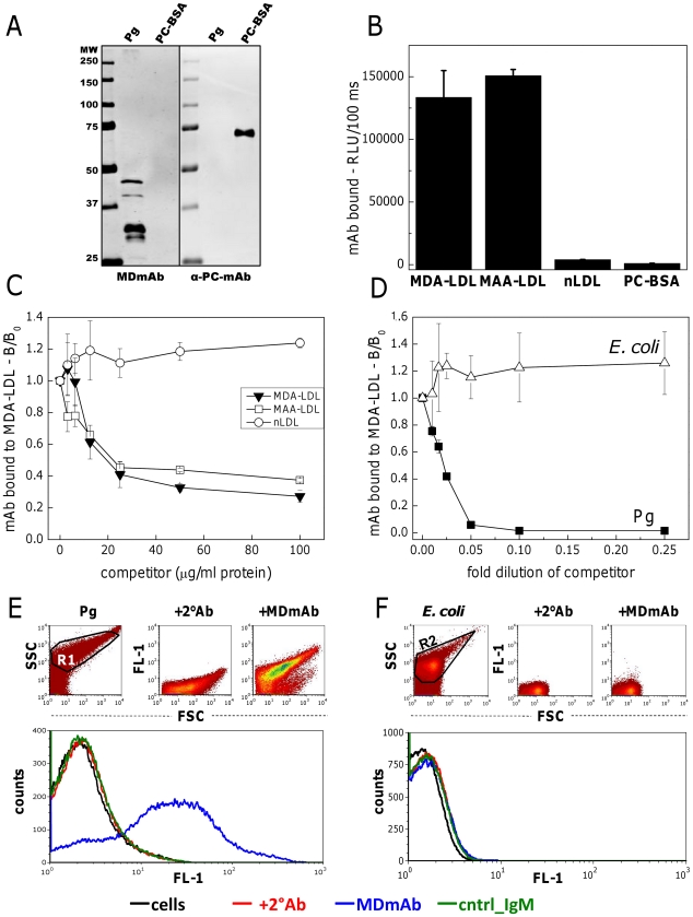
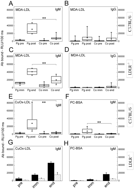
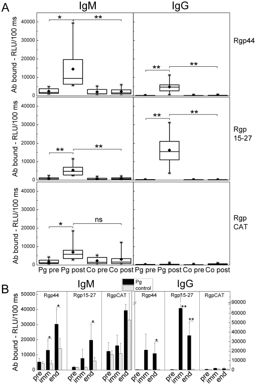
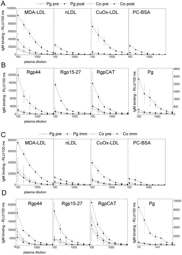
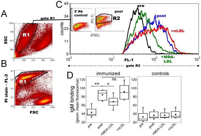
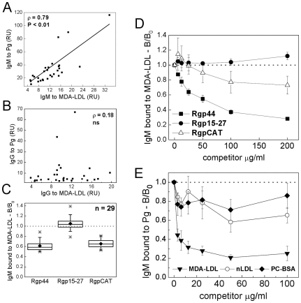
Methods and results: Mouse monoclonal IgM (MDmAb) specific for MDA-LDL recognized epitopes on P. gingivalis on flow cytometry and chemiluminescence immunoassays. Immunization of C57BL/6 mice with P. gingivalis induced IgM, but not IgG, immune response to MDA-LDL and apoptotic cells. Immunization of LDLR(-/-) mice with P. gingivalis induced IgM, but not IgG, immune response to MDA-LDL and diminished aortic lipid deposition. On Western blot MDmAb bound to P. gingivalis fragments identified as arginine-specific gingipain (Rgp) by mass spectrometry. Recombinant domains of Rgp produced in E. coli were devoid of phosphocholine epitopes but contained epitopes recognized by MDmAb and human serum IgM. Serum IgM levels to P. gingivalis were associated with anti-MDA-LDL levels in humans.

Conclusion: Gingipain of P. gingivalis is recognized by natural IgM and shares molecular identity with epitopes on MDA-LDL. These findings suggest a role for natural antibodies in the pathogenesis of two related inflammatory diseases, atherosclerosis and periodontitis.


Conflict of interest statement

Competing Interests: The authors have declared that no competing interests exist.

Figures

Figure 1
Figure 1. Cross-reactive epitopes on MDA-LDL and bacteria.
Binding of anti-MDA-LDL-IgM (MDmAb) and anti-PC-IgM control antibody (α-PC-mAb) to P. gingivalis (Pg) and PC-conjugated bovine serum albumin (PC-BSA) on Western blot (A). Binding of MDmAb to MDA- and MAA-modified and native LDL (nLDL) and PC-BSA using direct binding (B) and competitive (C) chemiluminescence immunoassays. Specific binding of MDmAb to P. gingivalis (Pg) and E. coli was tested with competitive chemiluminescence immunoassay (D). Bacterial suspensions were adjusted to an absorbance of 0.15 at 580 nm with PBS and further diluted as indicated. B/B0 indicates the ratio of IgM binding with and without a competitor. RLU, relative light unit. Binding of MDmAb and isotype control (cntrl_IgM) to P. gingivalis (E) and E. coli (F) in native conditions was tested with flow cytometry. Fluorescence (FL-1) of the cells with the secondary antibody, +2°Ab (red), MDmAb (blue) and isotype control (green).
Figure 2
Figure 2. Identification of P. gingivalis epitopes for anti-MDA-LDL-IgM.
A) Schematic presentation of arg-gingipain (RgpA) functional domains cloned and produced in a recombinant system . B) Proteins of P. gingivalis were separated on SDS-PAGE (Pg, gel). Fragments recognized by MDmAb (45, 40 and 32 kDa, black arrows) were identified by mass spectrometry as arginine-specific gingipain or hemagglutinin A of P. gingivalis. Figures S1, S2, S3 contain the Mascot results of the database searches and the MSMS spectrum showing the matching amino acids in the peptide sequence. Three domains of the recombinant arg-specific gingipain, RgpCAT, Rgp44 and Rgp15–27 were produced in E. coli and analyzed for recognition by MDmAb and anti-PC-IgM control antibody (α-PC-mAb). C) Specific binding of MDmAb to recombinant gingipain domains Rgp15–27, Rgp44 and RgpCAT was tested with a competitive immunoassay. D) Reciprocally, soluble MDA-LDL, nLDL and PC-BSA were used as competitors for MDmAb binding to Rgp44. B/B0 indicates the ratio of IgM binding with and without a competitor. MW, molecular weight. PC-BSA, phosphocholine-conjugated bovine serum albumin.
Figure 3
Figure 3. Mouse plasma IgM and IgG binding to MDA-LDL after immunization with P. gingivalis.
C57BL/6 mice were immunized with heat-killed P. gingivalis ATCC33277 (Pg; n = 8) and controls received saline (Co; n = 8). Plasma IgM (A) and IgG (B) to MDA-LDL before (pre) and after immunization (post) were determined with chemiluminescence immunoassay. Each C57BL/6 plasma sample (1∶500) was measured in duplicate and an average for each individual was calculated. LDLR−/− mice were immunized with killed P. gingivalis (3 strains mixed) (Pg; n = 7) and controls received PBS (Co; n = 8). Plasma IgM (C) and IgG (D) to MDA-LDL after the second booster immunization (imm) and after the HFD (end) were determined. Each LDLR−/− plasma sample (1∶1000) was measured in duplicate and an average for each individual in two repeated assays was calculated. Additionally, mouse plasma IgM binding to CuOx-LDL (E, G) and PC-BSA (F, H) was determined. For C57BL/6 mice (E, F) this was done similarly as described for panel A. Plasma samples of LDLR−/− mice (G, H) were pooled between three or four mice (1∶1000) for a single assay, in which the mean ± SD within a group is shown.
Figure 4
Figure 4. Antibodies to recombinant gingipain in P. gingivalis immunized mice.
Recombinant proteins of the arginine specific gingipain protease of P. gingivalis were produced in E.coli: two proteins in the hemagglutinin/adhesion domain, Rgp44 and Rgp15–27, and the catalytic domain, RgpCAT, which were used in chemiluminescence immunoassay to determine mouse plasma (1∶500) IgM and IgG binding in P. gingivalis immunized (Pg) and control (Co) groups in both A) C57BL/6 and B) LDLR−/− mice. A) For C57BL/6 immunized and control mice the samples (n = 8 each) were determined as duplicate before (pre) and after (post) immunization. Box-plots represent the distribution of the means of the sample duplicates. B) Two plasma samples within the group were pooled for immunized (black bars) and control (hatched bars) LDLR−/− mice and measured in duplicate. Samples were collected before (pre), after the second booster immunization (imm) and at the end of HFD (end). Columns represent the mean ± SD of pooled samples in each group. **P<0.01 and *P<0.05.
Figure 5
Figure 5. Dilution curves of mouse plasma IgM binding to antigens.
A, B) C57BL/6 mice were immunized with heat-killed P. gingivalis ATCC33277 (Pg) and controls (Co) received saline. The plasma was diluted 1∶100–1∶6400 and IgM binding to MDA-LDL, CuOx-LDL, native LDL, PC-BSA (A), P. gingivalis and recombinant gingipain domains Rgp44, Rgp15–27 and RgpCAT (B) were determined before (pre) and after (post) immunization with chemiluminescence immunoassay. Mean ± SD for two samples is shown. C, D) LDLR−/− mice were immunized with killed P. gingivalis (3 strains mixed) (Pg) and controls (Co) received PBS. Pooled plasma from two mice was used for each dilution curve, and mean ± SD for two dilution curves is shown. IgM binding to MDA-LDL, CuOx-LDL, native LDL, PC-BSA (C), P. gingivalis and recombinant gingipain domain Rgp44, Rgp15–27 and RgpCAT (D) were determined before (pre) and after the second booster immunization (imm) as described.
Figure 6
Figure 6. Mouse plasma IgM binding to apoptotic T lymphocytes after P. gingivalis immunization.
C57BL/6 mice were immunized with heat-killed Pg and controls received sterile saline (n = 8 per group). Mouse plasma (1∶70) IgM binding to UV-irradiated Jurkat T cells was measured with flow cytometry. A, B) Apoptotic T cell population (R1) was verified with propidium iodide (PI) staining. C) Plasma IgM binding in gate R2 of preimmune (black) and postimmune (blue) plasma samples, and competition of IgM binding with 250 µg/ml MDA-LDL (green) or native LDL (red). Inset plots (in Fig. 6C) represent the secondary antibody control (2°Ab control) and plasma IgM binding to apoptotic cells in a Pg-immunized mouse (post). D) IgM binding to Jurkat cells was determined for each mouse in Pg-immunized and control group as geometric mean value in R2 subtracted by the 2°Ab control. Box-plot graphs represent the distribution of sample means calculated for two repeated assays. **P<0.01 and *P<0.05.
Figure 7
Figure 7. Quantification of atherosclerosis in LDLR−/− mice immunized with P. gingivalis.
LDLR−/− mice (n = 7) were immunized without adjuvant with killed P. gingivalis (3 strains mixed) (Pg) followed by high fat diet (HFD). Controls (PBS, n = 8) received PBS. A) The extent of atherosclerotic plaque development was determined after HFD by en face analysis of the Sudan IV -stained aortas. B) Lesions at the aortic origin were measured on histological sections as percentage of plaque area in the aorta cross-sectional area. Representative pictures of aortas and cross-sections are shown for each group. * P<0.05.
Figure 8
Figure 8. Association between human serum IgM to P. gingivalis and MDA-LDL, and competitive binding with recombinant gingipain domains.
Sera from 29 healthy adults were analyzed for IgM (A) and IgG (B) binding to Pg and MDA-LDL by chemiluminescence immunoassay. Associations between antibody levels were analyzed with Spearman rank correlation test. Human sera were pre-incubated with recombinant gingipain domains Rgp44, Rgp15–27, RgpCAT (C, D) in a competitive immunoassay detecting IgM binding to immobilized MDA-LDL. The ratio of serum IgM binding (B/B0) to MDA-LDL with and without competitor (175 µg/ml) in 29 human serum samples (C) and dose-dependent competition assays of one sample (D). Reciprocal competition assay was performed to analyze human serum IgM binding to Pg antigen competed with MDA-LDL, nLDL and PC-BSA in a representative sample (E). RU, relative units.

Similar articles

Cited by

References

    1. Ross R. Atherosclerosis–an inflammatory disease. N Engl J Med. 1999;340:115–126. - PubMed
    1. Epstein SE, Zhu J, Burnett MS, Zhou YF, Vercellotti G, et al. Infection and atherosclerosis: Potential roles of pathogen burden and molecular mimicry. Arterioscler Thromb Vasc Biol. 2000;20:1417–1420. - PubMed
    1. Yusuf S, Hawken S, Ounpuu S, Dans T, Avezum A, et al. Effect of potentially modifiable risk factors associated with myocardial infarction in 52 countries (the INTERHEART study): Case-control study. Lancet. 2004;364:937–952. - PubMed
    1. Georges JL, Rupprecht HJ, Blankenberg S, Poirier O, Bickel C, et al. Impact of pathogen burden in patients with coronary artery disease in relation to systemic inflammation and variation in genes encoding cytokines. Am J Cardiol. 2003;92:515–521. - PubMed
    1. Esterbauer H, Schaur RJ, Zollner H. Chemistry and biochemistry of 4-hydroxynonenal, malonaldehyde and related aldehydes. Free Radic Biol Med. 1991;11:81–128. - PubMed

Publication types