. 2012;7(4):e34961.
doi: 10.1371/journal.pone.0034961. Epub 2012 Apr 4.

A family-wide RT-PCR assay for detection of paramyxoviruses and application to a large-scale surveillance study

Affiliations

A family-wide RT-PCR assay for detection of paramyxoviruses and application to a large-scale surveillance study

Sander van Boheemen et al. PLoS One. 2012.

Abstract

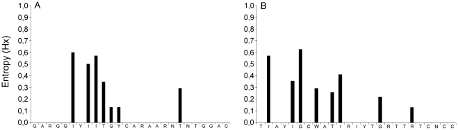
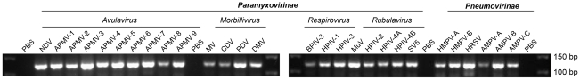
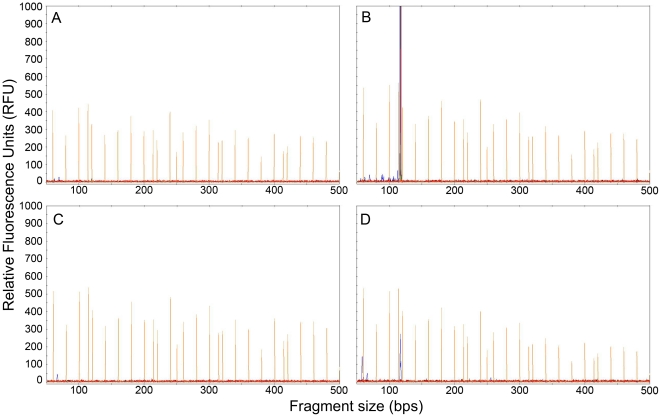
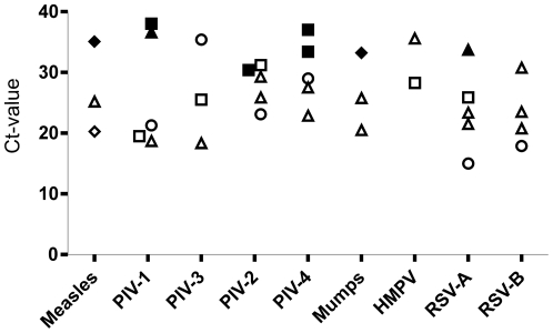
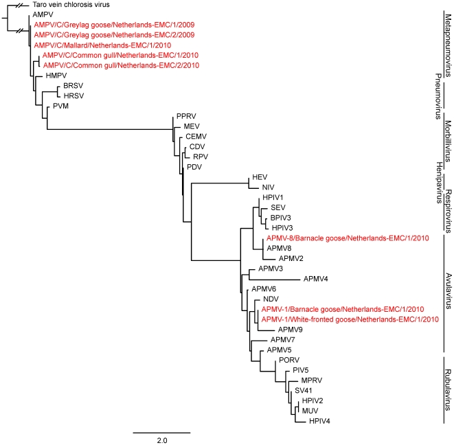
Family-wide molecular diagnostic assays are valuable tools for initial identification of viruses during outbreaks and to limit costs of surveillance studies. Recent discoveries of paramyxoviruses have called for such assay that is able to detect all known and unknown paramyxoviruses in one round of PCR amplification. We have developed a RT-PCR assay consisting of a single degenerate primer set, able to detect all members of the Paramyxoviridae family including all virus genera within the subfamilies Paramyxovirinae and Pneumovirinae. Primers anneal to domain III of the polymerase gene, with the 3' end of the reverse primer annealing to the conserved motif GDNQ, which is proposed to be the active site for nucleotide polymerization. The assay was fully optimized and was shown to indeed detect all available paramyxoviruses tested. Clinical specimens from hospitalized patients that tested positive for known paramyxoviruses in conventional assays were also detected with the novel family-wide test. A high-throughput fluorescence-based RT-PCR version of the assay was developed for screening large numbers of specimens. A large number of samples collected from wild birds was tested, resulting in the detection of avian paramyxoviruses type 1 in both barnacle and white-fronted geese, and type 8 in barnacle geese. Avian metapneumovirus type C was found for the first time in Europe in mallards, greylag geese and common gulls. The single round family-wide RT-PCR assay described here is a useful tool for the detection of known and unknown paramyxoviruses, and screening of large sample collections from humans and animals.


Conflict of interest statement

Competing Interests: Author Dr. Fouchier acts as an editor for PLoS ONE. This does not alter the authors' adherence to all the PLoS ONE policies on sharing data and materials.

Figures

Figure 1
Figure 1. Entropy plots of primer-annealing sites in human and animal paramyxovirus sequences available from Genbank.
Primer positions are given in the 5′ to 3′ direction. The sequences recognized by oligonucleotides PMX1 and PMX2 were compared to representative paramyxovirus genera and species sequences (N = 33), and their heterogeneities are displayed in panels A and B, respectively. The degree of heterogeneity was expressed as entropy as defined by Shannon: H (1) −∑f(b, 1) ln [f(b, 1)], where H (1) is the uncertainty at position 1, b represents a residue out of the allowed choices for the sequence in question (A, C, G, T, –), and f(b, 1) is the frequency at which residue b is found at position 1 , .
Figure 2
Figure 2. Detection of a wide range of paramyxoviruses by a single RT-PCR reaction.
RNA was isolated from the indicated virus stocks and, after cDNA synthesis, used for PCR analysis and subsequent agarose gel electrophoresis. Genomic sequences of twenty-eight paramyxoviruses from six genera were amplified by PCR using the PMX1/PMX2 primer pair. PBS indicates phosphate-buffered saline used as negative control in the entire procedure.
Figure 3
Figure 3. Fragment analysis plots of L gene fragments amplified by RT-PCR.
The orange peaks represent LIZ-600 size standards. The size standard peaks are 60, 80, 100, 114, 120, 140, 160, 180, 200, 214, 220, 240, 250, 260, 280, 300, 314, 320, 340, 360, 380, 400, 414, 420, 440, 460, 480, 500, 514, 520, 540, 560, 580 and 600 bps. The blue peaks are PCR products amplified by PMX1 and PMX2, where PMX1 is labelled with the 6-FAM fluorescent dye. A and C represent avian samples in which no paramyxoviruses were found. B and D show peaks around 121 nucleotides. Blue peaks in B and D represent APMV-1 and AMPV-C respectively. The relative fluorescence units are dependent on the amount of amplicon after RT-PCR.
Figure 4
Figure 4. Evaluation of the pan-paramyxovirus RT-PCR assay for virus detection in 35 clinical samples with Taqman-confirmed human paramyxoviruses.
Virus loads, as measured by Ct-values in virus specific Taqman assays, are plotted against paramyxovirus species (x-axis). Shapes of symbols indicate the type of clinical specimen (▵; oral samples, □; nose samples, ◊; lung samples, ⋄; other samples). Non-filled shapes represent samples that tested positive in fragment analysis, while filled shapes represent samples that tested negative.
Figure 5
Figure 5. Maximum likelihood (ML) phylogenetic tree based on a 70 nt L-gene fragment for 33 different Paramyxovirus species.
An L-gene fragment of the rhabdovirus Taro vein chlorosis virus (AY674964) was used as outgroup. Sequenced in black were obtained from Genbank while sequences in red were obtained from the avian surveillance study reported here. The length of the branch of Taro vein chlorosis virus as an outgroup is broken, and therefore does not represent its genetic distance to the rest of the tree. Accession numbers used for the phylogenetic tree were: NC003443, EU627591, NC009489, NC002200, NC009640, NC006430, NC006428, EU338414, EU403085, FJ177514, GU206351, NC003043, FJ231524, FJ215863, EU910942, NC002617, NC002161, C003461, NC001796, NC001552; NC001906, NC002728, NC001921, NC005283, NC001498, NC006383, Y09630, NC006296, NC001989, NC001781, NC006579, NC007652, NC004148.

References

    1. Lamb RA, Parks GD. Paramyxoviridae. In: Fields BN, Knipe DM, Howley PM, editors. Fields virology. 5th ed. Philadelphia: Wolters Kluwer/Lippincott Williams & Wilkins; 2007. pp. 1449–1496.
    1. Tregoning JS, Schwarze J. Respiratory viral infections in infants: causes, clinical symptoms, virology, and immunology. Clin Microbiol Rev. 2010;23:74–98. - PMC - PubMed
    1. Murray K, Selleck P, Hooper P, Hyatt A, Gould A, et al. A morbillivirus that caused fatal disease in horses and humans. Science. 1995;268:94–97. - PubMed
    1. Chua KB, Goh KJ, Wong KT, Kamarulzaman A, Tan PS, et al. Fatal encephalitis due to Nipah virus among pig-farmers in Malaysia. Lancet. 1999;354:1257–1259. - PubMed
    1. Chua KB, Bellini WJ, Rota PA, Harcourt BH, Tamin A, et al. Nipah virus: a recently emergent deadly paramyxovirus. Science. 2000;288:1432–1435. - PubMed

Publication types

MeSH terms