. 2012;7(4):e35036.
doi: 10.1371/journal.pone.0035036. Epub 2012 Apr 4.

Macrophage-associated mesenchymal stem cells assume an activated, migratory, pro-inflammatory phenotype with increased IL-6 and CXCL10 secretion

Affiliations

Macrophage-associated mesenchymal stem cells assume an activated, migratory, pro-inflammatory phenotype with increased IL-6 and CXCL10 secretion

Kevin Anton et al. PLoS One. 2012.

Abstract

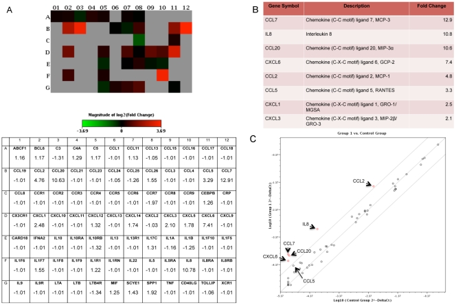
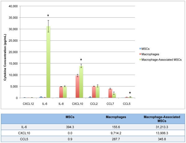
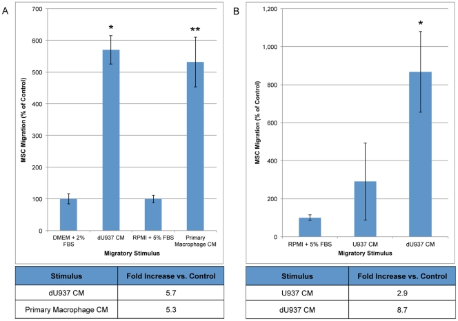
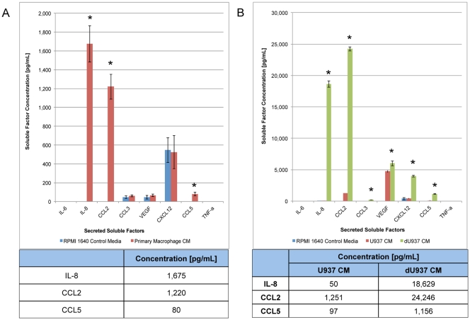
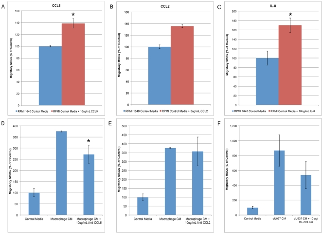
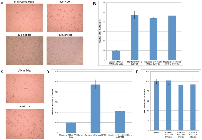
Mesenchymal stem cells (MSCs) exhibit tropism for sites of tissue injury and tumors. However, the influence of the microenvironment on MSC phenotype and localization remains incompletely characterized. In this study, we begin to define a macrophage-induced MSC phenotype. These MSCs secrete interleukin-6 (IL-6), CCL5, and interferon gamma-induced protein-10 (CXCL10) and exhibit increased mobility in response to multiple soluble factors produced by macrophages including IL-8, CCL2, and CCL5. The pro-migratory phenotype is dependent on activation of a c-Jun N-terminal kinase (JNK) pathway. This work begins to identify the influence of macrophages on MSC biology. These interactions are likely to play an important role in the tissue inflammatory response and may provide insight into the migratory potential of MSCs in inflammation and tissue injury.


Conflict of interest statement

Competing Interests: The authors have declared that no competing interests exist.

Figures

Figure 1
Figure 1. Inflammatory cytokine gene expression by MSCs is affected by macrophage-secreted soluble factors.
MSCs exposed to macrophage CM for 24 hours express an altered inflammatory cytokine gene expression profile. Analysis of 84 key genes mediating the inflammatory response was conducted using RT-PCR generating a heat map expressing log-fold changes. Gray boxes represent genes that were undetected in either the control MSCs or the macrophage CM-stimulated MSCs (A). When stimulated by macrophage CM, MSCs increase the expression of inflammatory factors such as, CCL7, IL-8, CCL20, CXCL6, CCL2, CCL5, CXCL1, and CXCL3. The changes ranged from 2.1 to 12.9-fold (B). These data can also be visualized in a scatter plot, showing gene up-regulation as points scattered into the upper left quadrant (C).
Figure 2
Figure 2. Macrophage-secreted soluble factors induce IL-6 and CXCL10 secretion by MSCs.
Macrophages and MSCs have distinct cytokine secretion profiles that are variable and significantly dependent on the soluble factors and cells in their local environment. Both macrophages and MSCs are highly plastic and assume unique phenotypes when activated by paracrine factors. MSCs cultured in macrophage-conditioned medium for 24 hours assume a pro-inflammatory phenotype with increased secretion of IL-6 and CXCL10. Resting MSCs secrete 394 pg/mL and an undetectable amount of IL-6 and CXCL10, respectively. Upon activation by macrophage-conditioned medium, MSCs increased their secretion of IL-6 to 31,213 pg/mL and CXCL10 to 13,906 pg/mL. This represents a 56-fold increase in IL-6 (p<0.0001) and a 1.4-fold increase in CXCL10 (p<0.003). In addition, MSCs minimally increased the secretion of CCL5 by 1.2-fold from 287 pg/mL to 345 pg/mL (p <0.02).
Figure 3
Figure 3. Macrophages induce MSC migration through the release of soluble factors.
To determine the chemotactic potential of macrophages for MSCs the CM from primary culture macrophages and the dU937 macrophage cell line was harvested and used as the stimulus in a standard Boyden Chamber migration assay. Following an 18-hour migration, MSCs show increased motility and migration towards macrophage-like CM and primary macrophage CM when compared with control medium. Compared to control levels, the number of migrating MSCs is 5.7-fold greater towards dU937 CM (*p<0.003, n = 3) and 5.3-fold greater towards primary macrophage CM (**p<0.01, n = 3), respectively (A). A separate experiment was conducted to characterize the ability of dU937 macrophages alto induce MSC chemotaxis. When compared to control, the soluble factors secreted by the dU937 macrophage cell line induce an 8.7-fold increase in MSC migration while U937-secreted factors increased MSC migration 2.9-fold (B). dU937 macrophage cells induce a significantly higher amount of MSC migration than the undifferentiated cells (*p<0.03, n = 3).
Figure 4
Figure 4. Identification of soluble factors produced by macrophages.
Unstimulated macrophages generated from human peripheral blood monocytes secrete IL-8, CCL2, and CCL5 (A) and do not secrete detectable amounts of IL-6 or MIP-1α. dU937 macrophages secrete increased levels of IL-8, CCL2, CCL20, CCL5, VEGF, and CXCL12 when compared with both control medium and undifferentiated U937 CM (B). All samples were analyzed in triplicate. *p<0.05.
Figure 5
Figure 5. Stimulation of mesenchymal stem cells by CCL5, CCL2, and IL-8 secretion induces migration.
To determine the role of macrophage-secreted CCL5, CCL2 (MCP-1), and IL-8 in MSC migration, the level of migration in response to recombinant proteins was analyzed. CCL5 (A), CCL2 (B), and IL-8 (C) significantly increased the number of migratory MSCs in a standard Boyden chamber chemotaxis assay. To further characterize the role of CCL5, CCL2, and IL-8 in macrophage-induced MSC migration, neutralizing antibodies targeting these factors were added to macrophage CM prior to migration. While each individual factor induced MSC migration, only CCL5 inhibition reduced MSC migration in response to macrophage CM (D, E, F).
Figure 6
Figure 6. Macrophage-secreted soluble factors induce MSC migration by activating the c-Jun NH2-terminal kinase (JNK) signaling pathway.
MSCs were pre-incubated for 1 hour in control medium or control medium containing 25 µM ERK or p38 inhibitor. Neither ERK nor p38 inhibition influenced MSC migration in response to dU937 CM (A, B). MSCs were incubated in 25 µM JNK inhibitor for 1 hour prior to migration assay. JNK inhibition decreased MSC migration by 55% (*p< 0.006, n = 3)(C, D). The MSCs were incubated with the inhibitors for 18 hours after which MSC viability was calculated using trypan blue staining. JNK, p38, and ERK inhibitors did not significantly decrease MSC viability (E).
Figure 7
Figure 7. Macrophages induce activation of the c-Jun NH2-terminal kinase pathway in MSCs.
To validate the macrophage-induced activation of the c-Jun NH2-terminal kinase (JNK) signaling pathway in MSCs, protein levels of phosphorylated downstream components were assessed by immunoblotting. Exposure to CM from dU937 macrophages induced activation of JNK1, 2, 3 after 15 minutes (A). An increase in the expression of phospho-c-Jun and decrease in the expression of ATF-2 was observed after a 30-minute treatment with dU937 CM (B). These results suggest that in addition to its control of cellular differentiation, proliferation, and apoptosis, the AP-1 transcription factors may also play a role in cellular chemotaxis (C).

References

    1. Spaeth E, Klopp A, Dembinski J, Andreeff M, Marini F. Inflammation and tumor microenvironments: defining the migratory itinerary of mesenchymal stem cells. Gene Ther. 2008;15:730–738. - PubMed
    1. Ryu CH, Park SH, Park SA, Kim SM, Lim JY, et al. Gene Therapy of Intracranial Glioma Using Interleukin 12-Secreting Human Umbilical Cord Blood-Derived Mesenchymal Stem Cells. Human gene therapy, 22(6) 2011;733–43 - PubMed
    1. Bak XY, Dang HL, Yang J, Ye K, Lee EX, et al. Human Embryonic Stem Cell-derived Mesenchymal Stem Cells as Cellular Delivery Vehicles for Prodrug Gene Therapy of Glioblastoma. Human gene therapy. 2011;22(11):1365–77. - PubMed
    1. Yang B, Wu X, Mao Y, Bao W, Gao L, et al. Dual-targeted antitumor effects against brainstem glioma by intravenous delivery of tumor necrosis factor-related, apoptosis-inducing, ligand-engineered human mesenchymal stem cells. Neurosurgery. 2009;65:610–624. - PubMed
    1. Varney ML, Johansson SL, Singh RK. Tumour-associated macrophage infiltration, neovascularization and aggressiveness in malignant melanoma: role of monocyte chemotactic protein-1 and vascular endothelial growth factor-A. Melanoma Res. 2005;15:417–425. - PubMed

Publication types

MeSH terms