. 2012;7(4):e35102.
doi: 10.1371/journal.pone.0035102. Epub 2012 Apr 9.

Lipid composition of the human eye: are red blood cells a good mirror of retinal and optic nerve fatty acids?

Affiliations

Lipid composition of the human eye: are red blood cells a good mirror of retinal and optic nerve fatty acids?

Niyazi Acar et al. PLoS One. 2012.

Abstract

Background: The assessment of blood lipids is very frequent in clinical research as it is assumed to reflect the lipid composition of peripheral tissues. Even well accepted such relationships have never been clearly established. This is particularly true in ophthalmology where the use of blood lipids has become very common following recent data linking lipid intake to ocular health and disease. In the present study, we wanted to determine in humans whether a lipidomic approach based on red blood cells could reveal associations between circulating and tissue lipid profiles. To check if the analytical sensitivity may be of importance in such analyses, we have used a double approach for lipidomics.

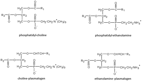
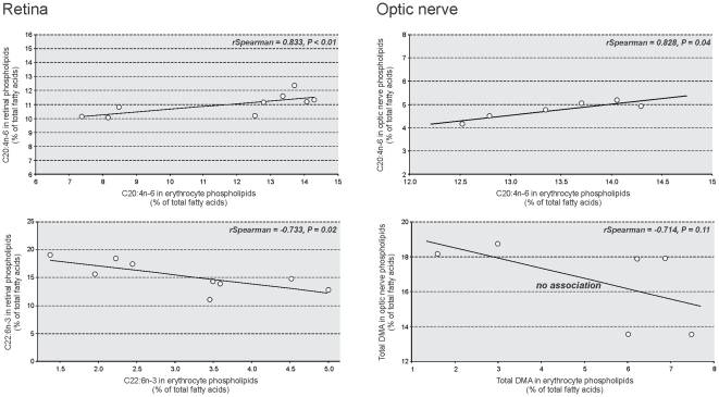
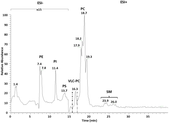
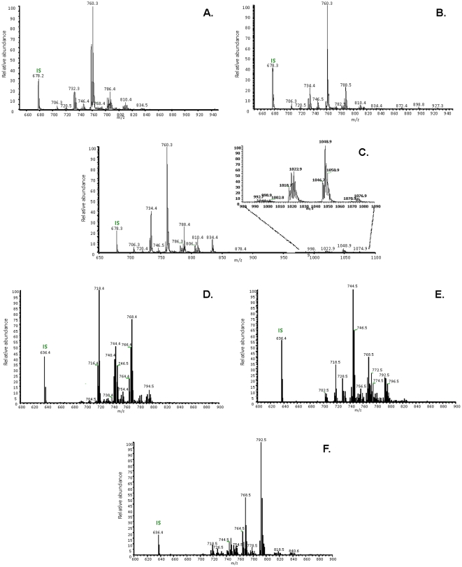
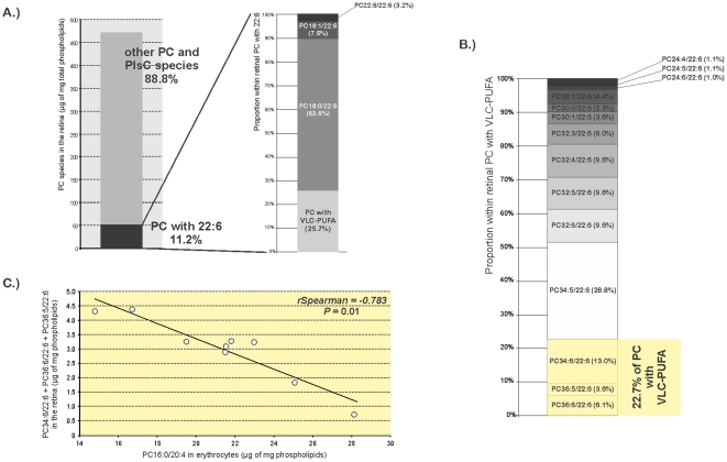
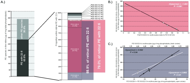
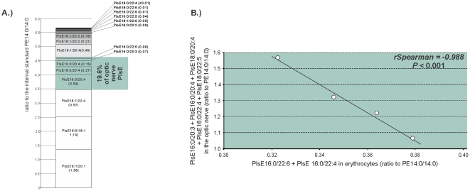
Methodology and principal findings: Red blood cells, retinas and optic nerves were collected from 9 human donors. The lipidomic analyses on tissues consisted in gas chromatography and liquid chromatography coupled to an electrospray ionization source-mass spectrometer (LC-ESI-MS). Gas chromatography did not reveal any relevant association between circulating and ocular fatty acids except for arachidonic acid whose circulating amounts were positively associated with its levels in the retina and in the optic nerve. In contrast, several significant associations emerged from LC-ESI-MS analyses. Particularly, lipid entities in red blood cells were positively or negatively associated with representative pools of retinal docosahexaenoic acid (DHA), retinal very-long chain polyunsaturated fatty acids (VLC-PUFA) or optic nerve plasmalogens.

Conclusions and significance: LC-ESI-MS is more appropriate than gas chromatography for lipidomics on red blood cells, and further extrapolation to ocular lipids. The several individual lipid species we have identified are good candidates to represent circulating biomarkers of ocular lipids. However, further investigation is needed before considering them as indexes of disease risk and before using them in clinical studies on optic nerve neuropathies or retinal diseases displaying photoreceptors degeneration.


Conflict of interest statement

Competing Interests: The authors have declared that no competing interests exist.

Figures

Figure 1
Figure 1. Structure of conventional phospholipids and plasmalogens.
Conventional phospholipids such as phosphatidyl-choline and phosphatidyl-etanolamine contain ester bonds in order to link R1 and R2 acyl- moieties at the sn-1 and sn-2 positions of glycerol, respectively. As for ethanolamine- and choline- plasmalogens, they have a vinyl ether bond at the sn-1 position of the glycerol backbone to link alkenyl- moieties and an ester bond at the sn-2 position to link acyl- residues.
Figure 2
Figure 2. Selected significant associations between erythrocyte and retinal or optic nerve lipids after gas chromatographic analyses.
Erythrocyte arachidonic acid (C20:4n-6) was positively associated with retinal C20:4n-6 (rSpearman = 0.833, P<0.01) and optic nerve C20:4n-6 (rSpearman = 0.828, P = 0.04). Erythrocyte docosahexaenoic acid (C22:6n-3, DHA) was negatively associated with retinal DHA (rSpearman = −0.733, P = 0.02) whereas no significant association emerged between erythrocyte and optic nerve levels of plasmalogens (evaluated by dimethylacetals, DMA).
Figure 3
Figure 3. LC-ESI-MS normal-phase chromatogram of the lipid extract from human retina.
The retention times of phosphatidyl-ethanolamine (PE), phosphatidyl-inositol (PI), phosphatidyl-serine (PS), phosphatidyl-choline (PC), sphingomyelin (SM), and lyso-phosphatidyl-choline (LPC) classes were of 7–8.5 min, 11–12 min, 12–14 min, 15.5–22 min, and 23–27.5 min, respectively. The mass spectrometer was operated under full scan in the negative ion mode from 0 to 15 min and in the positive ion mode from 15 min to 40 min.
Figure 4
Figure 4. Positive-ion HPLC-ESI-MS Mass spectra of total phosphatidyl-choline fraction collected from human neural retina.
A.) optic nerve (B.) and red blood cells (C.), by scanning for precursors at m/z 184 amu in the positive mode. Positive-ion HPLC-ESI-MS Mass spectra of total PE fraction collected from human neural retina D.), optic nerve (E.) and red blood cells (F.), using neutral loss scan at 141 amu in the positive mode.
Figure 5
Figure 5. Erythrocyte PC16:0/20:4 as a possible marker of a pool of retinal VLC-PUFA.
A): In the retina, VLC-PUFA accounted for about 25% of retinal PC species esterified to DHA, themself representing 11% of retinal total PC and PlsC. B): PC34:6/22:6, PC36:6/22:6, and PC36:5/22:6 were the longest and the most unsaturated VLC-PUFA in the retina. These three species accounted for 22.7% of total retinal VLC-PUFA. C) This pool of retinal VLC-PUFA was negatively associated with erythrocyte PC16:0/20:4 (rSpearman = −0.783, P = 0.01). Abbreviations of individual PC species are as follows: position on the glycerol backbone as shown as sn-1/sn-2 of the fatty alcohol radicals (abbreviated as number of carbons: number of double bonds).
Figure 6
Figure 6. Identification of circulating indexes of retinal PE esterified to DHA.
A) PE with DHA accounted for 63% of total retinal PE species. Within these entities, two different pools of molecules were of concern as they represented 79% and 89% of the total PE with 22:6. B) PlsE18:0/20:4 in erythrocytes was negatively associated to the first group represented by PE18:0/22:6+PE18:1/22:6+PE20:3/22:6 (rSpearman = −1.000, P<0.001). C) The second fraction of retinal PE with 22:6 represented by PE16:0/22:6+PE18:0/22:6+PE18:1/22:6 was positively associated to PE18:0/22:4 (black circles, rSpearman = 0.950, P = 0.04) and PE18:0/20:4+PE18:0/22:5 (open circles, rSpearman = −0.995, P = 0.01) in erythrocytes. Abbreviations of individual PE species are as follows: position on the glycerol backbone as shown as sn-1/sn-2 of the fatty alcohol radicals (abbreviated as number of carbons: number of double bonds).
Figure 7
Figure 7. PlsE16:0/22:6 and PlsE16:0/22:4 as indexes of a pool of optic nerve PlsE.
A) PlsE18:0/22:5, PlsE16:0/20:3, PlsE16:0/20:4, PlsE18:0/20:4, and PlsE16:0/22:4 represented 19% of optic nerve PlsE. B) This group of optic nerve PlsE was negatively associated to PlsE16:0/22:6 and PlsE16:0/22:4 in erythrocytes (rSpearman = −0.988, P<0.001). Abbreviations of individual PlsE species are as follows: position on the glycerol backbone as shown as sn-1/sn-2 of the fatty alcohol radicals (abbreviated as number of carbons: number of double bonds).

References

    1. Martinez M. Tissue levels of polyunsaturated fatty acids during early human development. J Pediatr. 1992;120:S129–138. - PubMed
    1. Martinez M, Ballabriga A, Gil-Gibernau JJ. Lipids of the developing human retina: I. Total fatty acids, plasmalogens, and fatty acid composition of ethanolamine and choline phosphoglycerides. J Neurosci Res. 1988;20:484–490. - PubMed
    1. Sastry PS. Lipids of nervous tissue: composition and metabolism. Prog Lipid Res. 1985;24:69–176. - PubMed
    1. Makrides M, Neumann MA, Byard RW, Simmer K, Gibson RA. Fatty acid composition of brain, retina, and erythrocytes in breast- and formula-fed infants. Am J Clin Nutr. 1994;60:189–194. - PubMed
    1. Das SK, Steen ME, McCullough MS, Bhattacharyya DK. Composition of lipids of bovine optic nerve. Lipids. 1978;13:679–684. - PubMed

Publication types