

Development of a novel preclinical pancreatic cancer research model: bioluminescence image-guided focal irradiation and tumor monitoring of orthotopic xenografts
- PMID: 22496923
- PMCID: PMC3323928
- DOI: 10.1593/tlo.11316
Development of a novel preclinical pancreatic cancer research model: bioluminescence image-guided focal irradiation and tumor monitoring of orthotopic xenografts
Abstract
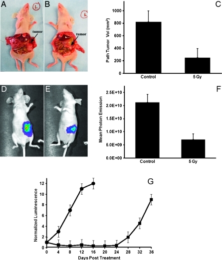
Purpose: We report on a novel preclinical pancreatic cancer research model that uses bioluminescence imaging (BLI)-guided irradiation of orthotopic xenograft tumors, sparing of surrounding normal tissues, and quantitative, noninvasive longitudinal assessment of treatment response.
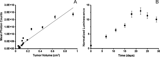
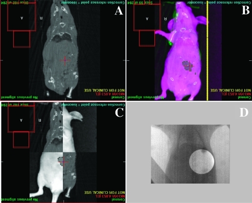
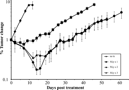
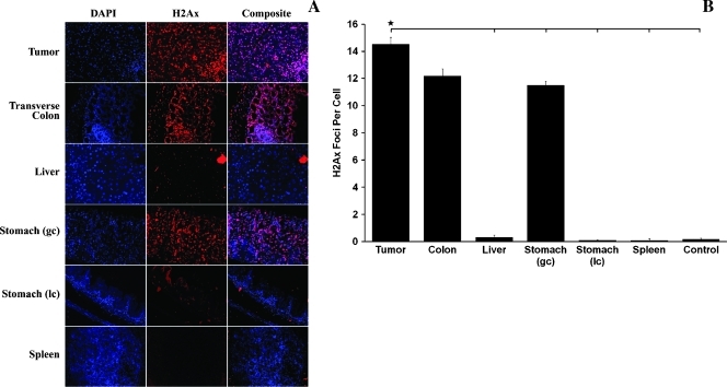
Materials and methods: Luciferase-expressing MiaPaCa-2 pancreatic carcinoma cells were orthotopically injected in nude mice. BLI was compared to pathologic tumor volume, and photon emission was assessed over time. BLI was correlated to positron emission tomography (PET)/computed tomography (CT) to estimate tumor dimensions. BLI and cone-beam CT (CBCT) were used to compare tumor centroid location and estimate setup error. BLI and CBCT fusion was performed to guide irradiation of tumors using the small animal radiation research platform (SARRP). DNA damage was assessed by γ-H2Ax staining. BLI was used to longitudinally monitor treatment response.
Results: Bioluminescence predicted tumor volume (R = 0.8984) and increased linearly as a function of time up to a 10-fold increase in tumor burden. BLI correlated with PET/CT and necropsy specimen in size (P < .05). Two-dimensional BLI centroid accuracy was 3.5 mm relative to CBCT. BLI-guided irradiated pancreatic tumors stained positively for γ-H2Ax, whereas surrounding normal tissues were spared. Longitudinal assessment of irradiated tumors with BLI revealed significant tumor growth delay of 20 days relative to controls.
Conclusions: We have successfully applied the SARRP to a bioluminescent, orthotopic preclinical pancreas cancer model to noninvasively: 1) allow the identification of tumor burden before therapy, 2) facilitate image-guided focal radiation therapy, and 3) allow normalization of tumor burden and longitudinal assessment of treatment response.
Figures
References
-
- Jemal A, Siegel R, Ward E, Hao Y, Xu J, Thun MJ. Cancer statistics, 2009. CA Cancer J Clin. 2009;59:225–249. - PubMed
-
- Herman JM, Swartz MJ, Hsu CC, Winter J, Pawlik TM, Sugar E, Robinson R, Laheru DA, Jaffee E, Hruban RH, et al. Analysis of fluorouracil-based adjuvant chemotherapy and radiation after pancreaticoduodenectomy for ductal adenocarcinoma of the pancreas: results of a large, prospectively collected database at the Johns Hopkins Hospital. J Clin Oncol. 2008;26:3503–3510. - PMC - PubMed
-
- Neoptolemos JP, Stocken DD, Friess H, Bassi C, Dunn JA, Hickey H, Beger H, Fernandez-Cruz L, Dervenis C, Lacaine F, et al. A randomized trial of chemoradiotherapy and chemotherapy after resection of pancreatic cancer. N Engl J Med. 2004;350:1200–1210. - PubMed
-
- Strimpakos A, Saif MW, Syrigos KN. Pancreatic cancer: from molecular pathogenesis to targeted therapy. Cancer Metastasis Rev. 2008;27:495–522. - PubMed
-
- Stojadinovic S, Low DA, Hope AJ, Vicic M, Deasy JO, Cui J, Khullar D, Parikh PJ, Malinowski KT, Izaguirre EW, et al. MicroRT-small animal conformal irradiator. Med Phys. 2007;34:4706–4716. - PubMed
LinkOut - more resources
Full Text Sources
Other Literature Sources