. 2011 Jul;2(3):125-141.
doi: 10.4161/self.17882. Epub 2011 Jul 1.

Cleavage of PGRP-LC receptor in the Drosophila IMD pathway in response to live bacterial infection in S2 cells

Affiliations

Cleavage of PGRP-LC receptor in the Drosophila IMD pathway in response to live bacterial infection in S2 cells

Rebecca L Schmidt et al. Self Nonself. 2011 Jul.

Abstract

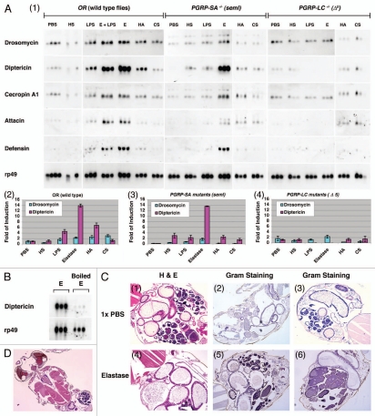
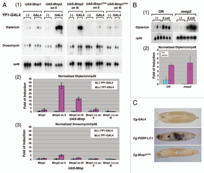
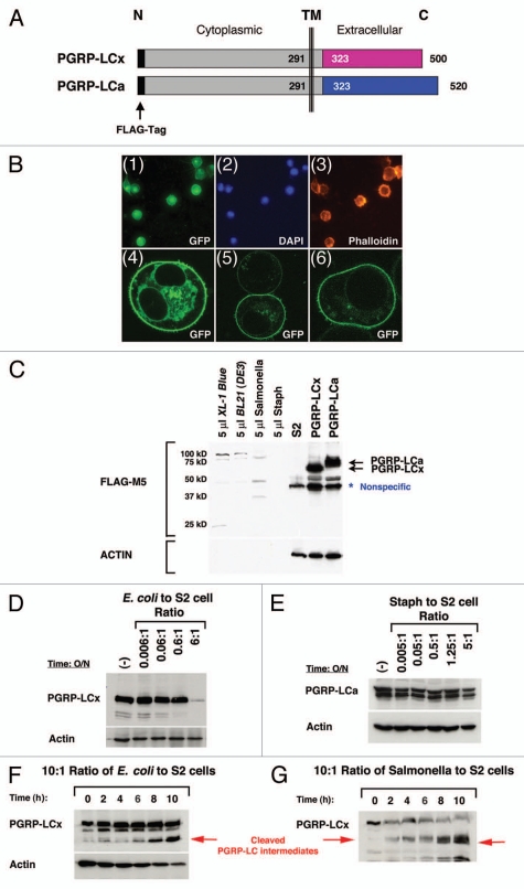
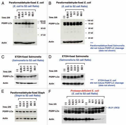
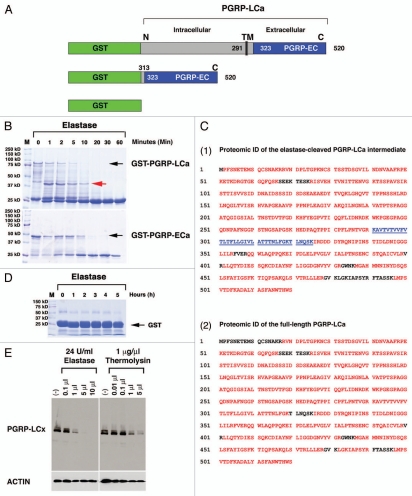
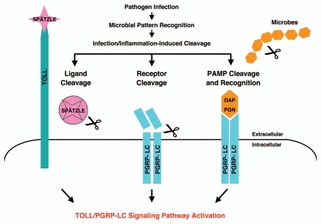
Drosophila responds to Gram-negative bacterial infection by activating the immune deficiency (IMD) pathway, leading to production of antimicrobial peptides (AMPs). As a receptor for the IMD pathway, peptidoglycan-recognition protein (PGRP), PGRP-LC is known to recognize and bind monomeric peptidoglycan (DAP-type PGN) through its PGRP ectodomain and in turn activate the IMD pathway. The questions remain how PGRP-LC is activated in response to pathogen infection to initiate the IMD signal transduction in Drosophila. Here we present evidence to show that proteases such as elastase and Mmp2 can also activate the IMD pathway but not the TOLL pathway. The elastase-dependent IMD activation requires the receptor PGRP-LC. Importantly, we find that live Salmonella/E. coli infection modulates PGRP-LC expression/receptor integrity and activates the IMD pathway while dead Salmonella/E. coli or protease-deficient E. coli do neither. Our results suggest an interesting possibility that Gram-negative pathogen infection may be partially monitored through the structural integrity of the receptor PGRP-LC via an infection-induced enzyme-based cleavage-mediated activation mechanism.


Figures

Figure 1
Figure 1
Elastase activates the IMD pathway through the receptor PGRP-LC. (A) Expression of antimicrobial peptides (AMP) was examined in triplicate in wild-type OR flies and mutant flies, PGRP-LC5 null) and PGRP-SA (seml), after treatment with sterile 1X PBS, elastase (3 Unit/ml), LPS (40 µg/ml), heparan sulfate (HS, 5 mg/ml), hyaluronic acid (HA , 20 mg/ml) or chondroitin sulfate (CS, 20 mg/ml) overnight. Northern blot analysis was performed using 50 µg total RNA extracted from the treated flies, and the blots were hybridized with 32P-labeled cDNA probes to antimicrobial peptides including Drosomycin, Diptericin, Cecropin A1, Attacin, and Defensin. rp49 mRNA was used as a control. No bacteria were added in these experiments to induce IMD activation. Film exposure is in the linear range. AMP production was examined by northern blots (panel 1). The relative levels of AMP expression, Diptericin/rp49 and Drosomycin/rp49, are shown in bar graphs in wild-type and mutant flies (panels 2, 3 and 4). Note that elastase was sufficient to induce robust IMD activation, whereas the saccharides did not. (B) AMP production in OR flies treated with elastase (3 U/ml) and heat-inactivated elastase (boiled at 100°C for 5 min) was examined. Note that the loss of elastase enzymatic activity is associated with loss of IMD activation. (C) PBS- and elastase-treated flies were embedded in paraffin blots. The intestinal morphologies of 1X PBS- and elastase-coated adult flies are shown. Hematoxylin and Eosin staining (H&E) is used to view adult abdominal morphology/cellular histology using serial paraffin sections (panels 1 and 4). Gram staining was used to detect for the loss of structural integrity of elastase-treated flies and the presence of commensal bacteria in internal organs post elastase treatment (panels 2, 3, 5 and 6). Note that there is no bacterial presence outside of the intestines of elastase-treated flies when compared with PBS-treated flies, demonstrating that the intestinal integrity remains intact at the microscopic level and no commensal bacteria leaked into the internal organs upon elastase treatment. (D) Section shows the general morphology of adult retina, brain, heart and muscles, demonstrating that paraffin section can be easily adapted for Drosophila research of host-pathogen interaction.
Figure 2
Figure 2
Expression of Drosophila matrix metalloproteinase, Mmp2, in fat bodies is specific in activating the IMD pathway in vivo. (A) Expression of Mmp2 in female adult fat bodies (YP1-GAL4) resulted in a marked increase in Diptericin mRNA production in the absence of bacterial infection. Northern blot was performed as described above. Two independent transgenic lines carrying Mmp1, Mmp1C93A or Mmp2 transgene inserted on either chromosome II or III were tested. No bacteria were used in these experiments to induce IMD activation. AMP production was examined by northern blots (panel 1). The relative levels of AMP expression, Diptericin/rp49 and Drosomycin/rp49, are quantified by bar graphs in MMP-expressing flies (panels 2 and 3). Note that Mmp2 expression induced robust IMD activation whereas the Mmp1 did not. (B) Mmp2 mutant larvae have a normal IMD response in response to E. coli infection. Two hundred wild-type and Mmp2 mutant larvae at the first and second instar stage were treated with or without E. coli for six hours. The northern analysis was performed as described (panel 1). The relative levels of AMP expression, Diptericin/rp49, in response to infection are quantified by bar graph. (C) Expression of Mmp1C93A under the control of Cg-GAL4 resulted in tissue histolysis (holes), internal tissue damage and subsequent lethality, suggesting that Mmp1C93A is an active MMP. However, no melanization reaction was observed. The expression of extracellular PGRP domain-deleted receptor, PGRP-LC-I, is known to drive melanization reaction in the absence of an infection, and it was used as a control.
Figure 3
Figure 3
Loss of PGRP-LC integrity in response to live E. coli/Salmonella infection in S2 cells. (A) A schematic illustration of the full-length receptors, PGRP-LCa/x, is shown. Identical intracellular and transmembrane (TM) domains are depicted as gray boxes and divergent extracellular PGRPa/x domains are depicted as pink and blue boxes. The FLAG tag (black bar) is added to the intracellular N-terminus of PGRP-LCa/x. This intracellular FLAG-tagged PGRP-LCa/x should be protected from any extracellular cleavage events and thus facilitate the detection of any receptor cleavage intermediates in response to pathogen infection by the western blot analyses. (B) PGRP-LC is a membrane receptor that is prominently expressed on the cell surface. (Panels 1–3) Stable S2 cell lines expressing FLAG-tagged PGRP-LC were established under the control of the inducible metallothionein promoter. Immunofluorescent staining shows that PGRP-LC is expressed on the membrane surface (panel 1). DAP I and Phalloidin were used as controls to stain nuclei (panel 2) and F-actin (panel 3). (Panels 4–6) Human cancer cells expressing EGFP-tagged PGRP-LC were established under the control of the CMV promoter. Three representative confocal images of the transfected cells are shown. PGRP-LC is clearly a membrane protein with some vesicles, ER/Golgi staining in the cytoplasm. (C)–(H) The expression of intracellular FLAG-tagged PGRP-LCx/a in S2 cells in response to live E. coli/Salmonella/Staph infection was determined by western blot analysis. (C) Expression of PGRP-LCx/a was established in stable S2 cell lines (two black arrows). S2 cells were used as negative control. A minimal amount of anti-FLAG-M5 cross-reactivity was detected with bacterial proteins [E. coli/Salmonella/Staph/BL21(DE3)] used in this study. A nonspecific band that cross-reacted with the anti-FLAG-M5 mAb was occasionally detected in untreated S2 cells. No receptor cleavage was observed under sterile condition. (D) Dosage-dependent cleavage of PGRP-LCx by live E. coli was examined after the cells were inoculated overnight with increasing amounts of E. coli. At the infection ratio of 5:1 (bacteria to S2 cells), PGRP-LC is cleaved. (E) No PGRP-LCa cleavage was detected under similar and higher bacterial conditions upon Staph (Gram-positive bacteria) infection overnight. (F) and (G) In order to capture the receptor cleavage intermediates, the S2 cells expressing PGRP-LC were subjected to a short time course of E. coli or Salmonella infection at the infection ratio of 10:1 (bacteria to S2 cells) for 0–10 h. Some PGRP-LC cleavage intermediates were readily detected (as marked by the red arrows). Anti-FLAG-M5 mAb was used to detect PGRP-LCa/x expression and Actin was used as a loading control.
Figure 4
Figure 4
PGRP-LC expression is not affected by the presence of high concentration of dead bacteria or protease-deficient E. coli. The expression of FLAG-tagged PGRP-LCa/x in S2 cells in response to dead E. coli/Salmonella/Staph and protease-deficient BL21(DE3) infection was determined by western blot analysis. (A), (B), (C), (D) and (E) None of the structurally intact but dead bacteria (either paraformaldehyde-fixed or ethanol-fixed E. coli, Salmonella or Staph) triggered any PGRP-LC cleavage at very high infection ratio of 50:1 (10-fold higher concentration than live bacterial infection). (F) A protease-deficient E. coli strain, BL21(DE3), was used to infect S2 cells stably expressing FLAG-tagged PGRP-LCa/x proteins. No significant receptor cleavage was detected at a high infection ratio of 60:1. Anti-FLAG-M5 mAb was used to detect PGRP-LCa/x expression and Actin was used as a loading control.
Figure 5
Figure 5
PGRP-LCa is an elastase substrate in vitro and a membrane-expressing PGRP-LCx can be cleaved by mammalian elastase and bacterial MMP in S2 cells. GSH-sepharose beads were used to affinity-purify full-length (FL) GST-tagged PGRP-LCa (named GST-PGRP-LCa), GST-tagged extracellular (EC) PGRPa domain (named GST-PGRP-LCa-EC) and GST proteins. GST protein is used as a negative control. (A) A schematic illustration of GST-tagged-PGRP-LCaFL/EC is shown. (B) The full-length GST-PGRP-LCaFL/EC fusion proteins are marked by black arrows. Both the full-length and extracellular fragment of PGRP-LCa can be readily cleaved by elastase in vitro. Equal aliquots (20 µl) of the GST-PGRP-LC fusion proteins were treated with 1 µl of elastase at different time points (1–60 min). The cleaved GST-PGRP-LCFL/EC intermediates were separated by SDS-PA GE and visualized by Coomassie Blue staining. A cleaved PGRP-LCa intermediate marked by the red arrow was sent for proteomic identification (ID) of the putative cleavage sites on PGRP-LC by elastase. (C) Proteomic ID results showed that the cleavage of PGRP-LCaFL/EC by elastase is rather nonspecific and elastase can completely cleave the extracellular PGRPa domain in vitro. (Panels 1 and 2) The proteomic identification results of PGRP-LCa cleavage intermediate as marked by the red arrow in Figure 5B (panel 1) and the full length PGRP-LCa (panel 2) are shown. The peptides identified and matched to the published PGRP-LCa amino acid sequence from the MS data are depicted in red. The near complete MS peptide coverage of the entire PGPLC-LCa coding sequence was detected in both the cleaved intermediate and the full-length receptor, indicating that the cleavage sites are rather nonspecific. (Panel 1) The transmembrane domain of PGRP-LCa that is located between 291–325 amino acids is not detected in the elastase-cleaved PGRP-LCa intermediate (marked by the underlined blue color). One possibility is that elastase may cut PGRP-LC near the TM domain between 291–325 Lysine (K) positions. Another possibility is that this hydrophobic TM peptide may be lost during sample preparation for MS ID in the elastase cleaved PGRP-LC intermediate. (D) GST protein was used as a negative control and treated with elastase for an extended period (6 h). No cleavage of GST protein was observed. (E) Stable S2 cell lines expressing intracellular FLAG-tagged PGRP-LCx were established under the control of the inducible metallothionein promoter. Dosage-dependent cleavage of PGRP-LCx by elastase (24 U/ml) or bacterial MMP, thermolysin (1 µg/ml) was examined after the cells were inoculated for 2 h with increasing amounts of proteases in serum-free medium under sterile conditions. The results showed that PGRP-LCx was readily accessible to elastase/thermolysin-mediated proteolysis. Anti-FLAG-M5 mAb was used to detect PGRP-LC expression and Actin was used as a loading control.
Figure 6
Figure 6
A model summarizing the infection-induced protease-dependent cleavage of innate immunity sensors/receptors in response to pathogenic infection and tissue damage in Drosophila A schematic illustration of the protease-dependent activation of Drosophila IMD and TOLL pathways is shown. It is well established that the TOLL ligand Spätzle is processed by an infection-activated serine protease cascade and that the cleaved Spätzle binds to TOLL and thereby activates the TOLL pathway. The receptor PGRP-LC can be activated by binding to bacterial elicitors (monomeric or polymeric DAP -PGN).,, To complement the well-established mechanism of innate immune activation via microbial pattern recognition (PAMP), we hypothesize that PGRP-LC may also be cleaved by infection-induced proteases released during pathogen-host antagonism. The structural integrity of the sentinel receptor PGRP-LC may constitute a “tissue well-being” signal. The infection-induced protease release may be a “danger/damage” signal that may help the host cells to detect pathogen infection and tissue injury.

References

    1. Lemaitre B, Hoffmann J. The host defense of Drosophila melanogaster. Annu Rev Immunol. 2007;25:697–743. doi: 10.1146/annurev.immunol.25.022106.141615. - DOI - PubMed
    1. Brennan CA, Anderson KV. Drosophila: the genetics of innate immune recognition and response. Annu Rev Immunol. 2004;22:457–483. doi: 10.1146/annurev.immunol.22.012703.104626. - DOI - PubMed
    1. Royet J, Reichhart JM, Hoffmann JA. Sensing and signaling during infection in Drosophila. Curr Opin Immunol. 2005;17:11–17. doi: 10.1016/j.coi.2004.12.002. - DOI - PubMed
    1. Levashina EA, Langley E, Green C, Gubb D, Ashburner M, Hoffmann JA, et al. Constitutive activation of toll-mediated antifungal defense in serpin-deficient Drosophila. Science. 1999;285:1917–1919. doi: 10.1126/science.285.5435.1917. - DOI - PubMed
    1. Michel T, Reichhart JM, Hoffmann JA, Royet J. Drosophila Toll is activated by Gram-positive bacteria through a circulating peptidoglycan recognition protein. Nature. 2001;414:756–759. doi: 10.1038/414756a. - DOI - PubMed