. 2012:2:359.
doi: 10.1038/srep00359. Epub 2012 Apr 11.

Therapeutic potential of HIV protease-activable CASP3

Affiliations

Therapeutic potential of HIV protease-activable CASP3

Kosuke Miyauchi et al. Sci Rep. 2012.

Abstract

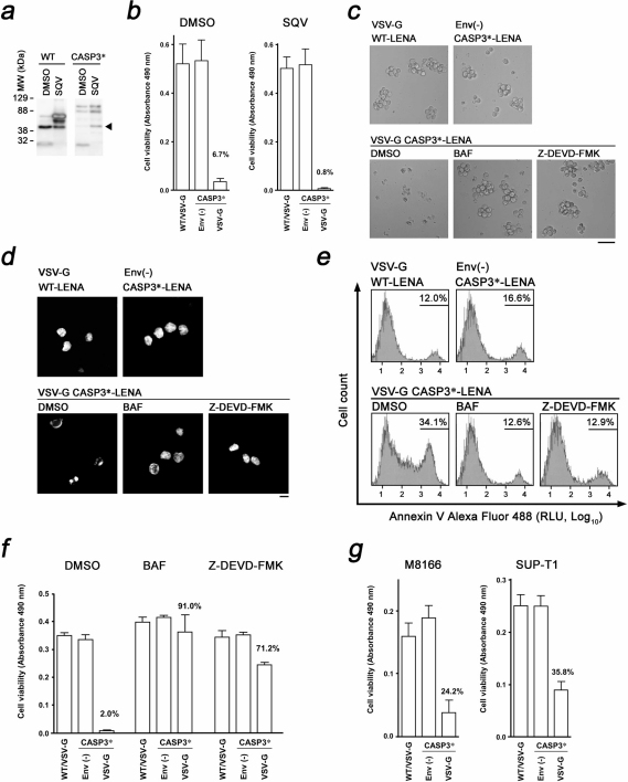
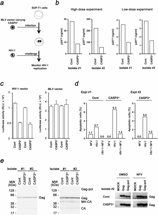
Development of a therapeutic application of CASP3/caspase 3/CPP32, an executor of apoptosis, has been challenging because regulation of its activation is complicated. This study aimed to inhibit cancer cell growth and human immunodeficiency virus type 1 (HIV-1) propagation through a CASP3 mutant, CASP3*, activable by HIV-1-encoded aspartate protease. Active CASP3* was delivered to leukemic cells using a protein transduction vehicle, the lentivirus-like nanoparticle (LENA), which should contain thousands of CASP3*-Gag protein molecules and release the activated CASP3* into the target cell cytoplasm. CASP3*-LENA induced apoptosis in various types of leukemic cells. In addition to being effective against leukemic cells, constitutive expression of CASP3* restricted HIV-1 propagation in SUP-T1 cells. The attenuation of HIV-1 replication in SUP-T1/CASP3* cells was attributed to the elimination of HIV-1-infected cells by apoptosis. These data suggest that CASP3* has therapeutic potential against both lymphoid malignancies and HIV-1 infection.


Figures

Figure 1
Figure 1. Construction, production and characterization of CASP3*-LENA.
(a) Genetic structure of CASP3 and the genetic modification to produce HIV-1 protease-activable CASP3*. The cellular (gray triangle, digesting D28-S29 and D175-S176 junctions) and HIV-1 (black triangle) protease proteolytic sites are indicated. The CASP3 D28 was replaced with amino acids 127–136 (VSQNYPIVQ) from HIV-1 Gag (according to the HXB2 coordinate). The amino acids 129–134 (QNYPIV) from HIV-1 Gag according to the HXB2 coordinate was inserted after CASP3 D175. HIV-1 protease targets the YP junction. The enzyme is active when the p17 and p12 subunits are dimerized. (b) Production of CASP3*-LENA and its mechanism of action. The CASP3* was placed at the 5’ end of wild type (WT) gag-pol to yield CASP3*-gag-pol. The proteolytic cleavage sites within Gag for HIV-1 protease and the proteolytic products are indicated MA, matrix; CA, capsid; and NC, nucleocapsid. The CASP3*-gag-pol and VSV-G expression vectors were co-transfected into 293T cells. Then, the VSV-G-encapsidated LENA containing activated CASP3* protein was produced in the culture supernatant. CASP3*-LENA enters cells via an endocytotic route, with the content released from endosomes at the site of membrane fusion. The action points of Bafilomycin A1 (BAF) and the CASP3 inhibitor Z-DEVD-FMK are indicated. (c) Expression of CASP3*-gag-pol and CASP3*-LENA production. The cell lysates (Cell) and culture supernatants (Sup) of 293T cells transfected with either wild type (WT) or CASP3*-gag-pol (CASP3*) expression vector were analyzed by Western Blot. The corresponding protein for each band is indicated. (d) Immunofluorescence assay showing the distribution of WT Gag or CASP3*-Gag (CASP3*) in 293T cells transfected with either WT or CASP3*-gag-pol (CASP3*) expression vector. Red and blue represent the anti-p24CA monoclonal antibody-stained signal and the Hoechst 33258-stained nucleus, respectively. Magnification, x630; scale bar, 10 µm. (e) Detection of CASP3 enzyme activity in purified CASP3*-LENA. CASP3*-LENA was produced either in the presence or absence of nelfinavir (NFV) and collected by ultracentrifugation over a 20% sucrose layer. DMSO was used as a control. The amount of CASP3*-LENA and the cleavage pattern of Gag were examined by Western blot (left). CASP3 enzyme activity was detected in the CASP3*-LENA lysate (right).
Figure 2
Figure 2. Induction of apoptosis in leukemic cells by CASP3*-LENA.
(a) Enhancement of WT- and CASP3*-LENA production by a low dose of saquinavir (SQV, 0.2 µM). DMSO was used as a solvent control. The arrowhead indicates a signal representing the MA-CA, indicating the release of CASP3* from CASP3*-Gag. (b) Quantitative analysis of MOLT-4 cell viability after LENA exposure. Cell viability was scored by colorimetric assay at 1d post-LENA exposure comparing WT-LENA encapsidated with VSV-G (WT/VSV-G), non-enveloped CASP3*-LENA (ENV(-)), and CASP3*-LENA encapsidated with VSV-G. The average and standard deviation from triplicated wells using LENA produced in the presence or absence of SQV are shown (SQV and DMSO, respectively). (c) Light microscopic observation of MOLT-4 cells treated with LENA produced in the presence of low-dose SQV. Cells were imaged at 1 d post-LENA exposure. Cells were treated with either Bafilomycin A1 (BAF) or the CASP3 inhibitor Z-DEVD-FMK. DMSO was used as a solvent control. Bar, 50 µm; magnification x100. (d) Nuclear morphology of MOLT-4 cells treated with LENA produced in the presence of low-dose SQV. Cells were imaged at 1 d post-LENA exposure after staining with Hoechst dye. Cells were treated with either BAF or Z-DEVD-FMK. DMSO was used as a solvent control. Bar, 10 µm; magnification x630. (e) Flow cytometric detection of apoptotic MOLT-4 cells. Cells were stained with Annexin V Alexa Fluor 488 at 6 h post-exposure to LENA produced in the presence of low dose nelfinavir (NFV, 0.2 µM). The live cell fraction was gated to analyze the early phase of apoptotic cell death. Cells were treated with either BAF or Z-DEVD-FMK. DMSO was used as a solvent control. RLU, relative light units. (f) Quantitative analysis of MOLT-4 cell viability after exposure to LENA produced in the presence of low-dose NFV (0.2 µM). Cell viability was scored at 1d post-LENA exposure. Cells were treated with either BAF or Z-DEVD-FMK. DMSO was used as a solvent control. (g) Quantitative analysis of M8166 and SUP-T1 cell viabilities as perfomed on MOLT-4 cells.
Figure 3
Figure 3. Inhibition of HIV-1 propagation by CASP3*.
(a) Schematic representation of the experimental approach. SUP-T1 cells were infected with MLV carrying the CASP3* to produce cells constitutively expressing CASP3*. These cells were infected with HIV-1, and the replication efficiency of HIV-1 was measured using viral p24CA antigen in the cell culture medium. (b) Inhibition of HIV-1NL4-3 replication in SUP-T1/CASP3* cells. Cells were exposed to high- or low-dose viral preparation yielding 25 ng or 1.3 ng of p24CA per 1 × 10⁵ cells, respectively. The viral p24CA concentration in the culture supernatant was measured at 6–9 d post-viral infection. Results shown were obtained from two independent isolates of SUP-T1/CASP3* and control cells. (c) Analysis of SUP-T1/CASP3* cell fate after HIV-1 entry. Luciferase activity of cells infected with either an HIV-1-based lentiviral vector or an MLV vector was measured at 4 d post-infection. RLU, relative light units. (d) Flow cytometric detection of apoptotic SUP-T1/Cont and SUP-T1/CASP3* cells by HIV-1NL4-3 infection. The percentage of Annexin V Alexa Fluor 488 positive cells was measured at 6 h post-nelfinavir (NFV) exposure, post-HIV-1 infection, and post-HIV-1 infection in the presence of 2 µM NFV, and subtracted from the percentage of Annexin V positive cells in untreated controls. The live cell fraction was gated according to scatter to analyze the early phase of apoptotic cell death. Data from two independent experiments are shown. For this experiment, cells from isolate #2 were used. (e) Analysis of SUP-T1/CASP3* cell fate upon HIV-1 production. The SUP-T1/CASP3* cells were transduced with HIV-1 Gag-pol or Gag by either MLV vector (left panel) or transfection (right panel), and the expression of transduced proteins was detected at 7 d post-gene transduction by Western blot. For the transfection experiment, cells were maintained in the presence of 0.5 µM NFV to detect the effect of Pol on cell survical.

References

    1. Cohen G. M. Caspases: the executioners of apoptosis. Biochem J. 326, 1–16 (1997). - PMC - PubMed
    1. Boatright K. M. & Salvesen G. S. Mechanisms of caspase activation. Curr Opin Cell Biol. 15, 725–31 (2003). - PubMed
    1. Yamabe K. et al. Cancer gene therapy using a pro-apoptotic gene, caspase-3. Gene Ther. 6, 1952–9 (1999). - PubMed
    1. Cam L., Boucquey A., Coulomb-L'hermine A., Weber A. & Horellou P. Gene transfer of constitutively active caspase-3 induces apoptosis in a human hepatoma cell line. J Gene Med. 7, 30–8 (2005). - PubMed
    1. Walters J. et al. A constitutively active and uninhibitable caspase-3 zymogen efficiently induces apoptosis. Biochem J. 424, 335–45 (2009). - PMC - PubMed

Publication types