. 2012 May 15;51(19):4062-71.
doi: 10.1021/bi300243z. Epub 2012 May 1.

Binding affects the tertiary and quaternary structures of the Shigella translocator protein IpaB and its chaperone IpgC

Affiliations

Binding affects the tertiary and quaternary structures of the Shigella translocator protein IpaB and its chaperone IpgC

Philip R Adam et al. Biochemistry. .

Abstract

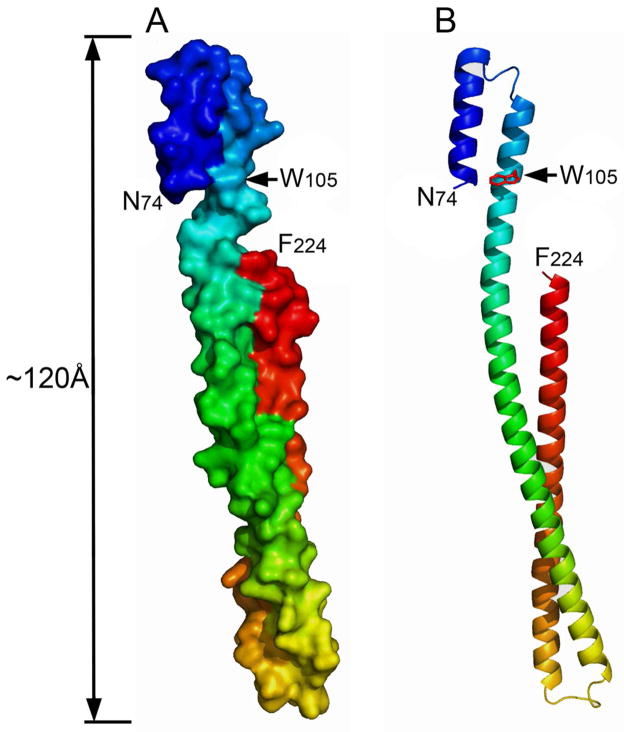
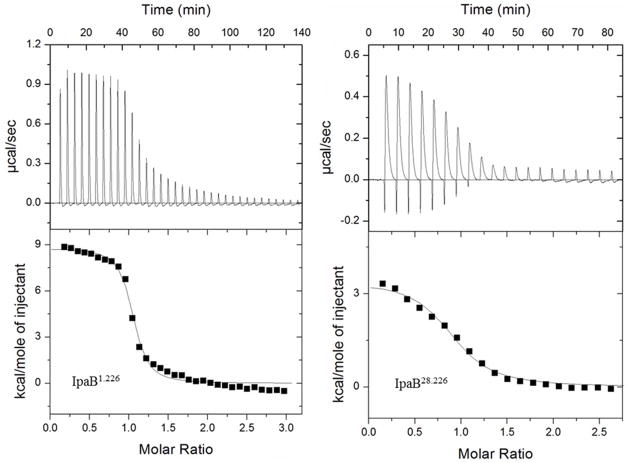
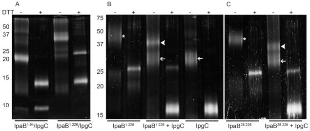
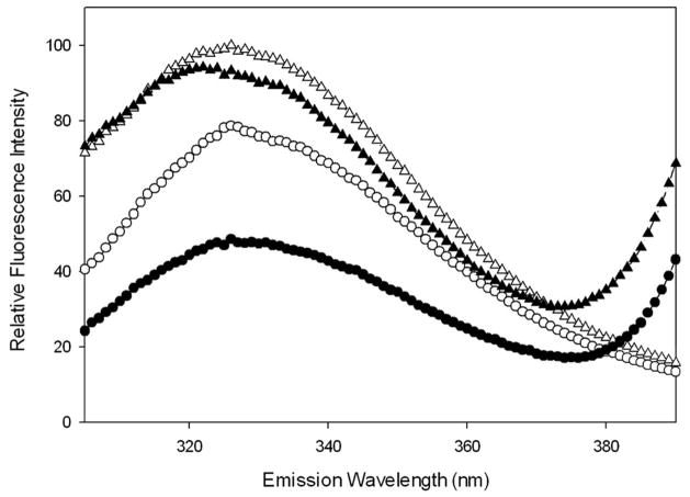
Shigella flexneri uses its type III secretion system (T3SS) to promote invasion of human intestinal epithelial cells as the first step in causing shigellosis, a life-threatening form of dysentery. The Shigella type III secretion apparatus (T3SA) consists of a basal body that spans the bacterial envelope and an exposed needle that injects effector proteins into target cells. The nascent Shigella T3SA needle is topped with a pentamer of the needle tip protein invasion plasmid antigen D (IpaD). Bile salts trigger recruitment of the first hydrophobic translocator protein, IpaB, to the tip complex where it senses contact with a host membrane. In the bacterial cytoplasm, IpaB exists in a complex with its chaperone IpgC. Several structures of IpgC have been determined, and we recently reported the 2.1 Å crystal structure of the N-terminal domain (IpaB(74.224)) of IpaB. Like IpgC, the IpaB N-terminal domain exists as a homodimer in solution. We now report that when the two are mixed, these homodimers dissociate and form heterodimers having a nanomolar dissociation constant. This is consistent with the equivalent complexes copurified after they had been co-expressed in Escherichia coli. Fluorescence data presented here also indicate that the N-terminal domain of IpaB possesses two regions that appear to contribute additively to chaperone binding. It is also likely that the N-terminus of IpaB adopts an alternative conformation as a result of chaperone binding. The importance of these findings within the functional context of these proteins is discussed.


Figures

Figure 1
Figure 1. The crystal structure of the N-terminal domain of IpaB shows 2.1 Å resolution for residues 74–224
The native Trp residue within this domain is indicated and Cys residues placed at the N or C terminus of IpaB1.226 and IpaB28.226 would not be visible in this structure, indicating that they are in unstructured regions within the otherwise stable N-terminal domain. Panel A shows a surface rendition of the structure (PDB 3U0C) and panel B shows a ribbon depiction for this region of IpaB (18). The position of Trp residue 105 is indicated with an arrow (W105) and the length of the entire structure is indicated.
Figure 2
Figure 2. Fluorescence polarization (FP) measurements for the binding of IpaB1.226 and IpaB28.226
Panel A) N-terminus FM-labeled IpaBN28.226 (open circles, R2=0.99) and IpaBN1.226 (closed circles, R2=0.94) or Panel B) C-terminus FM-labeled IpaBC28.226 (open circles, R2= 0.99) and IpaBC1.226 (closed circles, R2=0.99) were held at a constant concentration as increasing concentrations of IpgC were added. A single-site ligand binding saturation equation was fit to these data. Data shown are representative of three independent experiments with at least 3 technical replicates per experiment. Error bars represent standard deviation.
Figure 3
Figure 3. Isothermal titration calorimetry (ITC) measurements for IpgC and IpaB fragments
Titrations of IpgC with IpaB1.226 (left panel) and IpaB28.226 (right panel) as monitored by ITC. The upper panel of each section depicts the thermograms obtained during the course of each injection series. The corresponding integrated enthalpy changes (after background correction) are shown in each lower panel. A solid line is depicted in the lower panel that fits the data according to a single-site binding model. Experimentally derived thermodynamic values are listed in Table 3.
Figure 4
Figure 4. Chemical cross-linking analysis of the IpaB N-terminal domain and IpgC
Aliquots of protein (10μM) were exposed to 168 uM of the cross-linking agent DSP at room temperature for 30 min. Cross-linking was quenched by the addition of a third volume of 3x SDS-PAGE sample buffer. The presence or absence of DTT in the sample buffer is indicated at the top of the gels. In panel A, IpaB1.94 and IpaB1.226 were co-expressed and co-purified with IpgC followed by cross-linking as described above. In panel B, IpgC, and IpaB1.226 were expressed and purified independently. Proteins were cross-linked at IpaB:IpgC molar ratios of 0:1, 1:2, and 1:0. In panel C, IpaB28.226 was tested at 0 or a two-fold molar excess of IpgC. White arrowheads show the position of IpaB/IpgC homodimer, thin white arrows show the position of IpgC homodimers and the asterisk (*) indicates the position of IpaB N-terminal domain homodimers. Addition of DTT cleaves the DSP cross-linker, resulting in the formation of modified monomeric proteins.
Figure 5
Figure 5. Use of FRET to monitor intermolecular distances during the interaction of the IpaB N-terminal domain with IpgC
Alexa350-IpaBN1.226 (D/A) or IpaBN1.226 (D) was excited at 295 nm and fluorescence spectra were collected from 300–400 nm in the presence and absence of 1μM IpgC. Open circles, IpaBN1.226 donor only without IpgC; open triangles, IpaBN1.226 donor only with IpgC; closed circles, IpaBN1.226 donor plus acceptor without IpgC; and closed triangles, IpaBN1.226 donor with acceptor and IpgC present. The data shown are representative of the data used to obtain the FRET efficiency values given in Table 4. The fluorescence spectra shown here are normalized relative to IpaBN1.226 donor only in the presence of IpgC.

Similar articles

Cited by

References

    1. Mueller C, Broz P, Cornelis G. The type III secretion system tip complex and translocon. Mol Microbiol. 2008;68:1085–1095. - PubMed
    1. Cornelis GR. The type III secretion injectisome, a complex nanomachine for intracellular ‘toxin’ delivery. Biol Chem. 2010;391:745–751. - PubMed
    1. Schroeder GN, Hilbi H. Molecular pathogenesis of Shigella spp.: controlling host cell signaling, invasion, and death by type III secretion. Clin Microbiol Rev. 2008;21:134–156. - PMC - PubMed
    1. Espina M, Olive AJ, Kenjale R, Moore DS, Ausar SF, Kaminski RW, Oaks EV, Middaugh CR, Picking WD, Picking WL. IpaD Localizes to the Tip of the Type III Secretion System Needle of Shigella flexneri. Infect Immun. 2006;74:4391–4400. - PMC - PubMed
    1. Darboe N, Kenjale R, Picking WL, Picking WD, Middaugh CR. Physical characterization of MxiH and PrgI, the needle component of the type III secretion apparatus from Shigella and Salmonella. Protein Sci. 2006;15:543–552. - PMC - PubMed

Publication types

MeSH terms