. 2012 Jun 4;9(6):1599-611.
doi: 10.1021/mp2006054. Epub 2012 May 4.

Cationic PAMAM dendrimers disrupt key platelet functions

Affiliations

Cationic PAMAM dendrimers disrupt key platelet functions

Clinton F Jones et al. Mol Pharm. .

Abstract

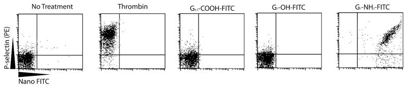
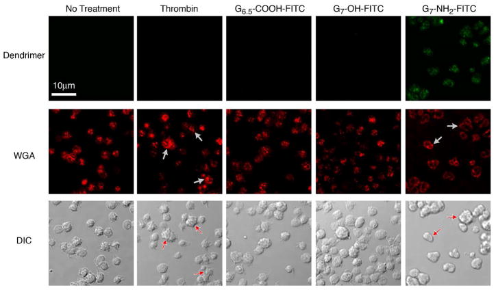
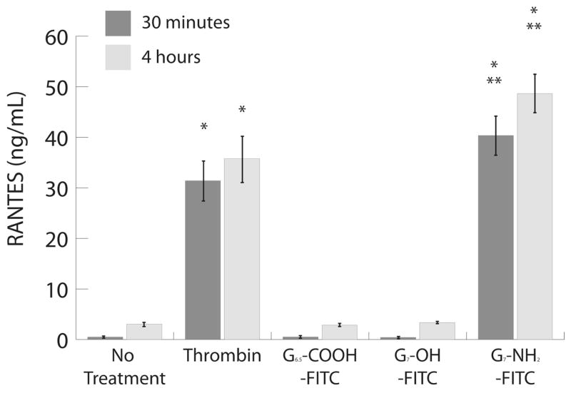
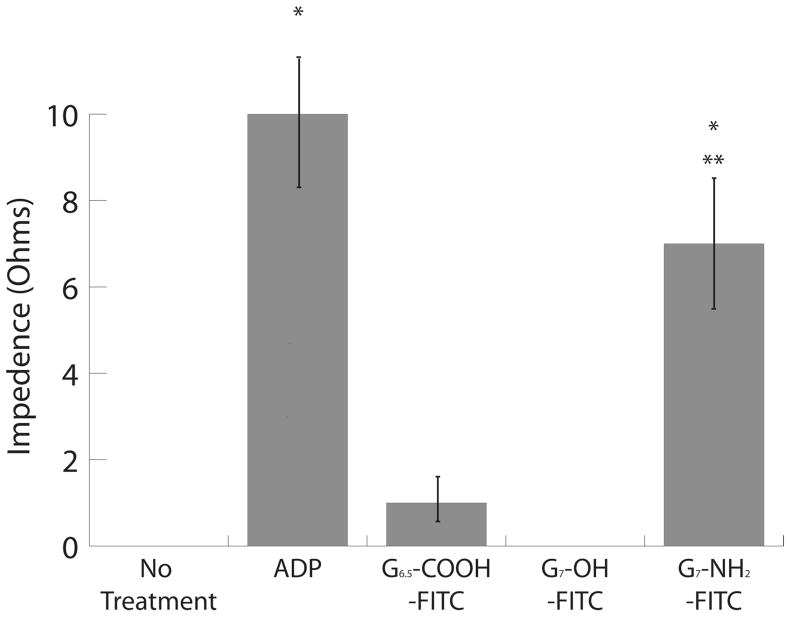
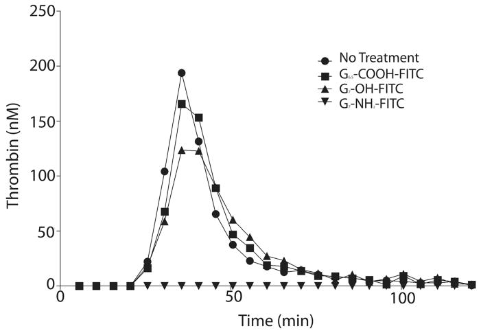
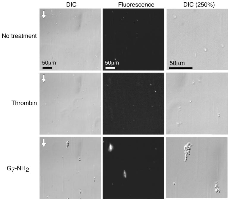
Poly(amidoamine) (PAMAM) dendrimers have been proposed for a variety of biomedical applications and are increasingly studied as model nanomaterials for such use. The dendritic structure features both modular synthetic control of molecular size and shape and presentation of multiple equivalent terminal groups. These properties make PAMAM dendrimers highly functionalizable, versatile single-molecule nanoparticles with a high degree of consistency and low polydispersity. Recent nanotoxicological studies showed that intravenous administration of amine-terminated PAMAM dendrimers to mice was lethal, causing a disseminated intravascular coagulation-like condition. To elucidate the mechanisms underlying this coagulopathy, in vitro assessments of platelet functions in contact with PAMAM dendrimers were undertaken. This study demonstrates that cationic G7 PAMAM dendrimers activate platelets and dramatically alter their morphology. These changes to platelet morphology and activation state substantially altered platelet function, including increased aggregation and adherence to surfaces. Surprisingly, dendrimer exposure also attenuated platelet-dependent thrombin generation, indicating that not all platelet functions remained intact. These findings provide additional insight into PAMAM dendrimer effects on blood components and underscore the necessity for further research on the effects and mechanisms of PAMAM-specific and general nanoparticle toxicity in blood.


Figures

Figure 1
Figure 1. G7-NH2-FITC dendrimers induce P-selectin expression on platelets (A-D)
Platelets were treated with saline, thrombin, or 100 μg/mL of different functionalized dendrimers for 30 minutes. Thrombin induces P-selectin expression while the G6.5-COOH-FITC and G7-OH-FITC dendrimer-treated platelets express significantly less P-selectin and negatively stain for FITC, indicating no dendrimer binding. In contrast, G7-NH2-FITC treated platelets express P-selectin and stain positively for FITC, suggesting dendrimer binding. The (*) indicates significantly different than no treatment (p<0.05). The (**) indicates significantly different than G6.5-COOH-FITC and G7-OH-FITC dendrimer (p<0.05). Flow diagrams are from a single experiment and are representative of three independent experiments (A). The data represent the mean ± SEM of at least three independent experiments.
Figure 2
Figure 2. G7-NH2-FITC dendrimers bind to and alter the morphology of platelets
Platelets were incubated with saline, thrombin (0.1 U/mL, final), or G6.5-COOH, G7-OH or G7-NH2-FITC dendrimer (100 μg/mL, final) for 45 minutes. Baseline platelets have few pseudopodia and intact granules compared to thrombin-activated platelets (A, second panel, see arrows). G6.5-COOH-FITC and G7-OH-FITC dendrimer-treated platelets have no apparent alterations in morphology, while G7-NH2-FITC dendrimer treated platelets have a ruffled exterior, loss of granule morphology (see arrows) and appear green, indicating G7-NH2-FITC binding to the platelet (A, right panels). Platelets were treated with saline, thrombin (0.1 U/mL, final) or G7-NH2-FITC dendrimer (100 μg/mL, final) and imaged using a JEOL CarryScope SEM (B). Images were taken at 2,500X, with a 20 mm working distance and a 20 kV accelerating voltage. Notice the increased number and size of platelet aggregates as well as the ruffled membrane morphology of the G7-NH2 dendrimer treated platelets (B, right panel).
Figure 3
Figure 3. Human platelets release alpha granules contents in response to G7-NH2-FITC dendrimers
Whole blood treated with G7-NH2-FITC dendrimers results in significant activation of platelets based on RANTES and PF4 release after thirty minutes and four hours (A,B). Purified platelets release RANTES in a dose- and time-dependent manner after treatment with G7-NH2-FITC, but not G6.5-COOH-FITC or G7-OH2-FITC (C,D). The (*) indicates significantly different compared to no treatment (p<0.05). The (**) indicates significantly different compared to G6.5-COOH-FITC and G7-OH-FITC dendrimer (p<0.05). The data represent the mean ± SEM of at least three independent experiments.
Figure 4
Figure 4. G7-NH2-FITC dendrimers induce platelet aggregation
Human whole blood (A) and PRP (B, C) were treated with G6.5-COOH-FITC, G7-OH-FITC, or G7-NH2-FITC, (100 μg/mL, final), saline, or ADP (5 μM, final). G7- NH2-FITC dendrimer treatment alone in significant platelet aggregation compared to no treatment (*, p<0.05) and G6.5-COOH-FITC and G7-OH-FITC (**, p<0.05) in whole blood. G7-NH2-FITC treatment significantly increases platelet aggregation in PRP (*, p<0.05) compared to no treatment as indicated by the increase in amplitude (B) and slope (C). The bars in panels A-C represent the mean ± SEM of three independent experiments.
Figure 5
Figure 5. G7-NH2-FITC dendrimers inhibit platelet-dependent thrombin generation in whole blood and PRP
Whole blood was treated with saline or dendrimer (100 μg/mL) for 30 minutes at 37°C. After 30 minutes, the whole blood was centrifuged at 150 × g for 20 minutes and the PRP was transferred to another tube. Thrombin generation was then measured using the PRP after addition of tissue factor, CaCl2, and a fluorogenic substrate. Thrombin generation was monitored with a fluorimeter for 120 minutes (A). G7-NH2-FITC dendrimers inhibited thrombin generation in whole blood, while the G6.5-COOH-FITC and G7-OH -FITC dendrimers had little effect. Dendrimer alone had no effect on the rate of thrombin cleavage of the fluorogenic substrate or on the measurement of fluorescence (data not shown). Whole blood was centrifuged for 150 × g for 20 minutes and PRP was transferred to another tube. PRP was treated with saline or dendrimer (100 μg/mL) for 30 minutes before thrombin generation was measured as described above. Similar to whole blood, G7-NH2-FITC dendrimers blunted thrombin generation in dendrimer treated PRP while G6.5-COOH-FITC and G7-OH -FITC dendrimers had no effect (B). Thrombin generation plots (A,B) depict one experiment and are representative of three independent experiments.
Figure 6
Figure 6. Platelets adhere faster and in greater numbers to fibrinogen under flow after G7-NH2 dendrimer treatment
Platelets were left untreated or stimulated with thrombin (0.5 U/mL) or G7-NH2 dendrimer (100 μg/mL) for five minutes and flowed at 200 s−1 for six minutes over Ibidi 0.4 VI plates pre-coated with 0.5 mg/mL fibrinogen. In some experiments, platelets were fluorescently labeled before treatment. Images were captured in real-time using an Olympus wide-field fluorescent microscope (IX-81 inverted microscope system) using an ORCA-ER monochrome CCD camera (frame rate 1Hz). Still images from the final frame are depicted in panel A from a single experiment, representative of twelve independent experiments. Images in panel B are still images from the final frame from one experiment, representative of six independent experiments. The flow direction is indicated by the arrow in the upper left hand corner (A, B). The DIC image has been enlarged by 250% in the right panels to show platelet morphology. The initial rate of platelet adhesion and average area covered by platelets were significantly (p<0.05) increased for dendrimer-treated platelets relative to untreated controls (C,D). In some experiments, platelets were pre-treated for one hour with abciximab (0.136 μM), a Fab fragment against αIIbβ3, before treatment with agonist. Abciximab-treated platelets showed reduced adhesion to fibrinogen, independent of agonist treatment (B–D). However, the aggregation behavior of platelets was not significantly different for abciximab-treated platelets stimulated with cationic dendrimer (E). The bars in panels C–E represent the mean ± SEM of at least six independent experiments. The (*) indicates significantly (p<0.05) different compared to no treatment. The (**) indicates significantly (p<0.05) different compared to non-abciximab-treated platelets.

Similar articles

Cited by

References

    1. Tomalia DA, Fréchet JMJ. Discovery of dendrimers and dendritic polymers: A brief historical perspective. Journal of Polymer Science Part A: Polymer Chemistry. 2002;40:2719–2728.
    1. Radu DR, Lai CY, Jeftinija K, Rowe EW, Jeftinija S, Lin VSY. A Polyamidoamine Dendrimer- Capped Mesoporous Silica Nanosphere-Based Gene Transfection Reagent. Journal of the American Chemical Society. 2004;126:13216–13217. - PubMed
    1. Bielinska AU, Yen A, Wu HL, Zahos KM, Sun R, Weiner ND, Baker JR, Jr, Roessler BJ. Application of membrane-based dendrimer/DNA complexes for solid phase transfection in vitro and in vivo. Biomaterials. 2000;21:877–887. - PubMed
    1. Svenson S, Tomalia DA. Dendrimers in biomedical applications--reflections on the field. Adv Drug Deliv Rev. 2005;57:2106–29. - PubMed
    1. Esfand R, Tomalia DA. Poly(amidoamine) (PAMAM) dendrimers: from biomimicry to drug delivery and biomedical applications. Drug Discov Today. 2001;6:427–436. - PubMed

Publication types