

Epileptic encephalitis: the role of the innate and adaptive immune system
- PMID: 22497613
- PMCID: PMC8029258
- DOI: 10.1111/j.1750-3639.2012.00580.x
Epileptic encephalitis: the role of the innate and adaptive immune system
Abstract
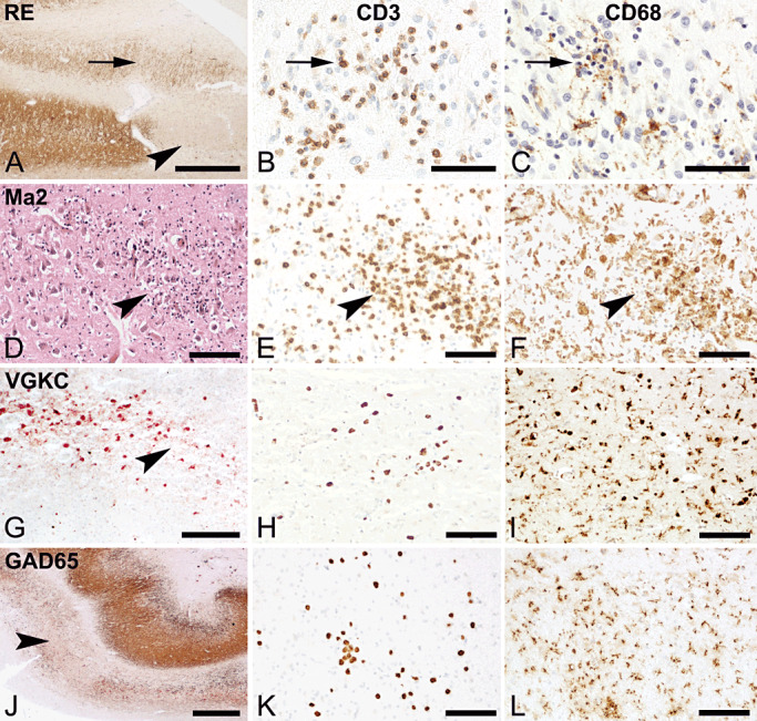
Seizures are a prominent clinical feature of encephalitis. Recent data suggest the adaptive as well as innate immune system to be involved directly in the pathomechanism of epileptogenesis. Cytotoxic T-cells and antibody-mediated complement activation are major components of the adaptive immune system, which can induce neurodegeneration, thereby probably contributing to epileptic encephalitis. The innate immune system operates via interleukin-1 and toll-like receptor-associated mechanisms and was shown to play a direct role in epileptogenesis. Here, we review neuropathology hallmarks of various encephalitis conditions such as Rasmussen encephalitis (RE) but also introduce the more recently discovered antibody-associated voltage-gated potassium channel complex (VGKC), N-methyl-D-aspartate receptor (NMDAR) or glutamic acid decarboxylase (GAD) 65 encephalitides. Neuropathological investigations are used to determine specific cellular components and molecular mechanisms used by the immune system to provoke neurodegeneration and to promote epileptogenesis. Based on recent findings, we propose concepts for the stratification of epileptic encephalitis. Knowledge of the role of the innate immunity has already translated into clinical treatment strategies and may help to discover novel drug targets for these epileptic disorders.
© 2012 The Authors; Brain Pathology © 2012 International Society of Neuropathology.
Figures
Similar articles
-
Immunity and Inflammation in Epilepsy.Cold Spring Harb Perspect Med. 2015 Dec 18;6(2):a022699. doi: 10.1101/cshperspect.a022699. Cold Spring Harb Perspect Med. 2015. PMID: 26684336 Free PMC article. Review.
-
Immunopathology of autoantibody-associated encephalitides: clues for pathogenesis.Brain. 2012 May;135(Pt 5):1622-38. doi: 10.1093/brain/aws082. Epub 2012 Apr 25. Brain. 2012. PMID: 22539258
-
Inflammation, ictogenesis, and epileptogenesis: An exploration through human disease.Epilepsia. 2021 Feb;62(2):303-324. doi: 10.1111/epi.16788. Epub 2020 Dec 14. Epilepsia. 2021. PMID: 33316111 Review.
-
Autoimmune epilepsy in children: case series and proposed guidelines for identification.Epilepsia. 2013 Jun;54(6):1036-45. doi: 10.1111/epi.12142. Epub 2013 Mar 28. Epilepsia. 2013. PMID: 23551014
-
Immune-mediated epilepsies.Epilepsia. 2011 May;52 Suppl 3(Suppl 3):5-11. doi: 10.1111/j.1528-1167.2011.03029.x. Epilepsia. 2011. PMID: 21542839 Free PMC article. Review.
Cited by
-
Therapeutic Efficacy of Lavandula dentata's Oil and Ethanol Extract in Regulation of the Neuroinflammation, Histopathological Alterations, Oxidative Stress, and Restoring Balance Treg Cells Expressing FoxP3+ in a Rat Model of Epilepsy.Pharmaceuticals (Basel). 2024 Dec 31;18(1):35. doi: 10.3390/ph18010035. Pharmaceuticals (Basel). 2024. PMID: 39861097 Free PMC article.
-
Clinical studies and anti-inflammatory mechanisms of treatments.Epilepsia. 2017 Jul;58 Suppl 3(Suppl 3):69-82. doi: 10.1111/epi.13779. Epilepsia. 2017. PMID: 28675558 Free PMC article.
-
Does brain inflammation mediate pathological outcomes in epilepsy?Adv Exp Med Biol. 2014;813:169-83. doi: 10.1007/978-94-017-8914-1_14. Adv Exp Med Biol. 2014. PMID: 25012376 Free PMC article. Review.
-
Pathomorphological Diagnostic Criteria for Focal Cortical Dysplasias and Other Common Epileptogenic Lesions-Review of the Literature.Diagnostics (Basel). 2023 Mar 31;13(7):1311. doi: 10.3390/diagnostics13071311. Diagnostics (Basel). 2023. PMID: 37046529 Free PMC article. Review.
-
Targeting inflammation as a therapeutic strategy for drug-resistant epilepsies: an update of new immune-modulating approaches.Hum Vaccin Immunother. 2014;10(4):868-75. doi: 10.4161/hv.28400. Epub 2014 Mar 7. Hum Vaccin Immunother. 2014. PMID: 24609096 Free PMC article. Review.
References
-
- Albert ML, Austin LM, Darnell RB (2000) Detection and treatment of activated T cells in the cerebrospinal fluid of patients with paraneoplastic cerebellar degeneration. Ann Neurol 47:9–17. - PubMed
-
- Andermann F (1991) Chronic Encephalitis and Epilepsy. Rasmussen's Syndrome. Butterworth‐Heinemann: Boston.
-
- Andrews PI, Dichter MA, Berkovic SF, Newton MR, McNamara JO (1996) Plasmapheresis in Rasmussen's encephalitis. Neurology 46:242–246. - PubMed
-
- Bauer J, Elger CE, Hans VH, Schramm J, Urbach H, Lassmann H, Bien CG (2007) Astrocytes are a specific immunological target in Rasmussen's encephalitis. Ann Neurol 62:67–80. - PubMed
