. 2013 Feb;39(2):157-65.
doi: 10.1111/j.1365-2990.2012.01274.x.

Nuclear carrier and RNA-binding proteins in frontotemporal lobar degeneration associated with fused in sarcoma (FUS) pathological changes

Affiliations

Nuclear carrier and RNA-binding proteins in frontotemporal lobar degeneration associated with fused in sarcoma (FUS) pathological changes

Y S Davidson et al. Neuropathol Appl Neurobiol. 2013 Feb.

Abstract

Aims: We aimed to investigate the role of the nuclear carrier and binding proteins, transportin 1 (TRN1) and transportin 2 (TRN2), TATA-binding protein-associated factor 15 (TAF15) and Ewing's sarcoma protein (EWS) in inclusion body formation in cases of frontotemporal lobar degeneration (FTLD) associated with fused in sarcoma protein (FTLD-FUS).

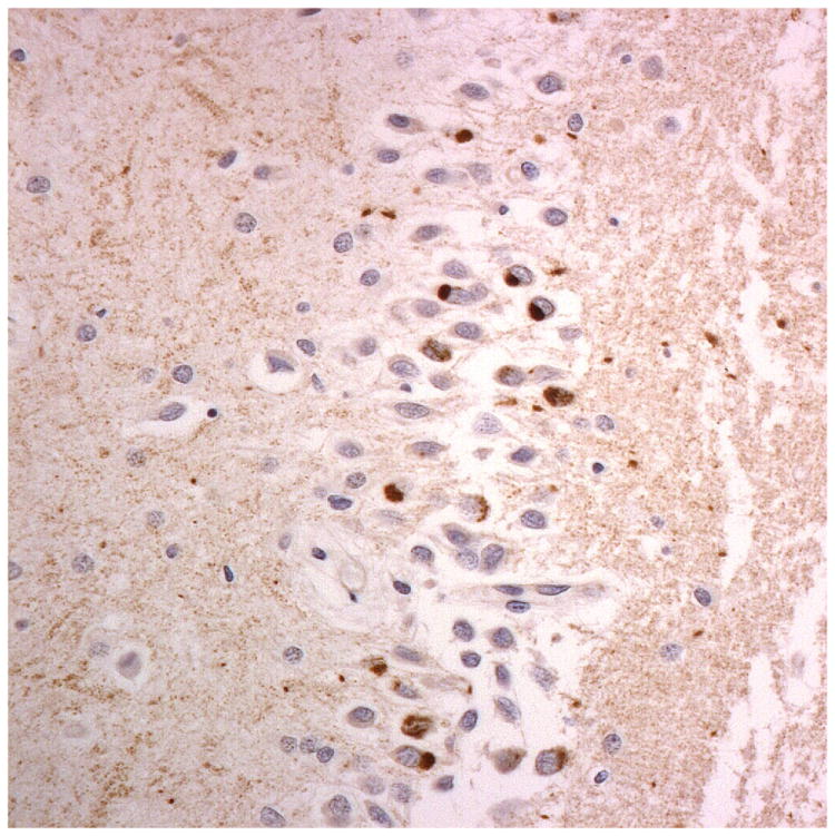
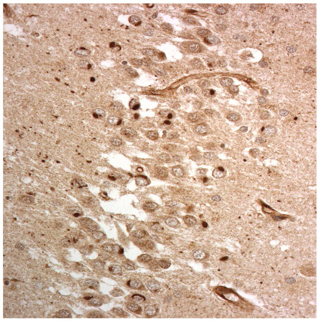
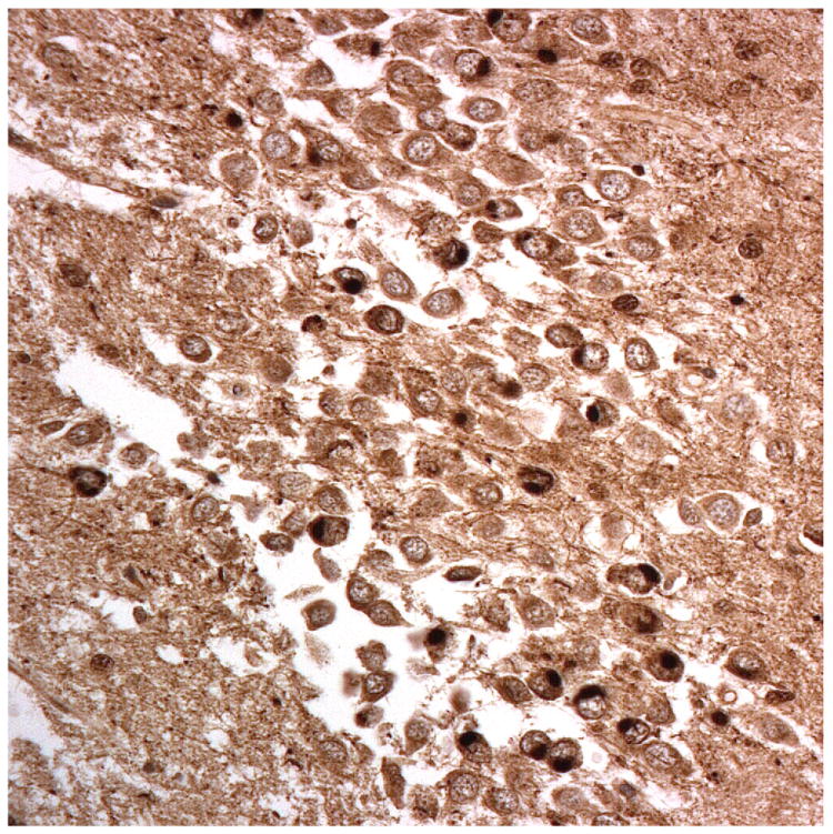
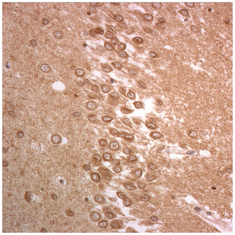
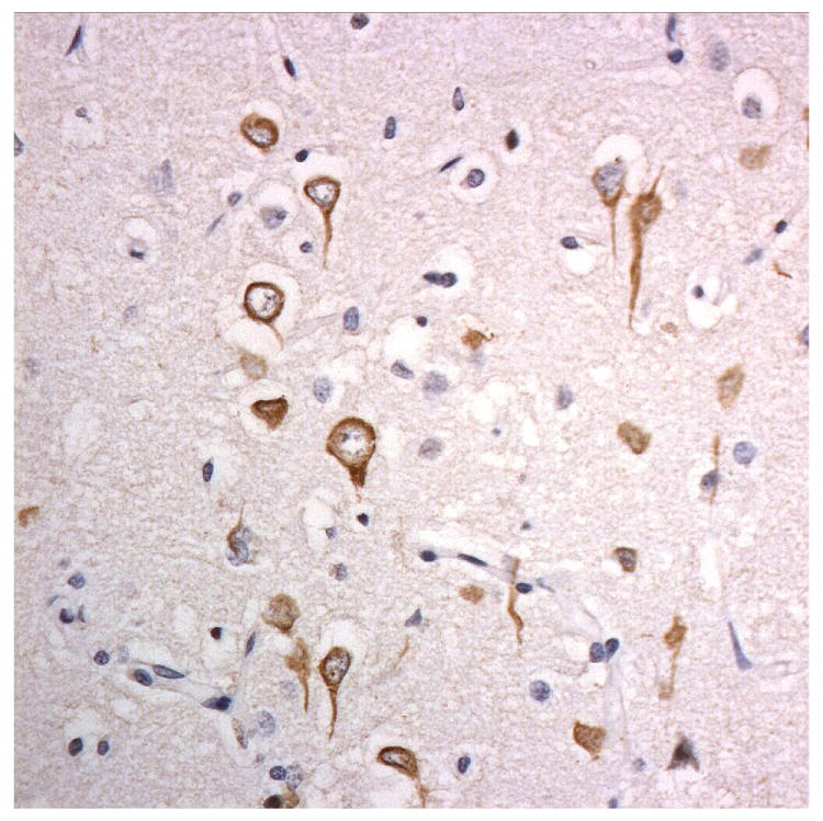
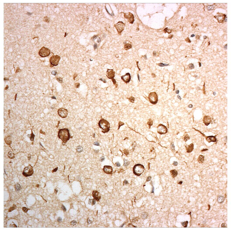
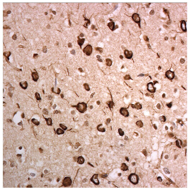
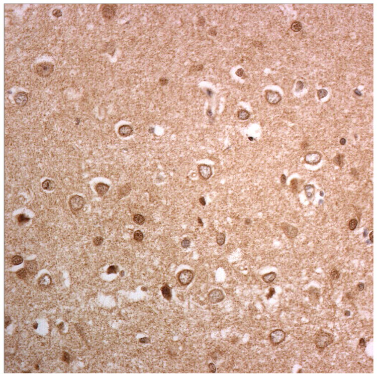
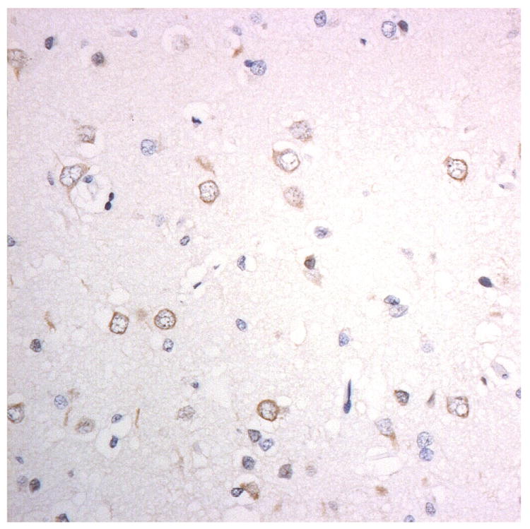
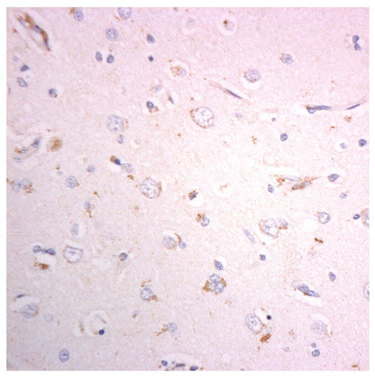
Methods: Eight cases of FTLD-FUS (five cases of atypical FTLD-U, two of neuronal intermediate filament inclusion body disease and one of basophilic inclusion body disease) were immunostained for FUS, TRN1, TRN2, TAF15 and EWS. Ten cases of FTLD associated with TDP-43 inclusions served as reference cases.

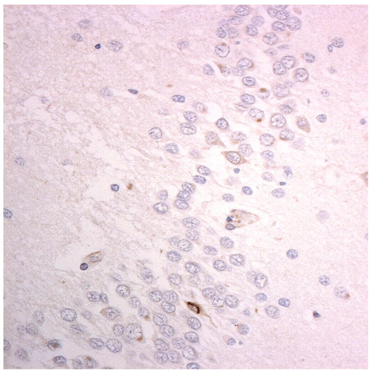
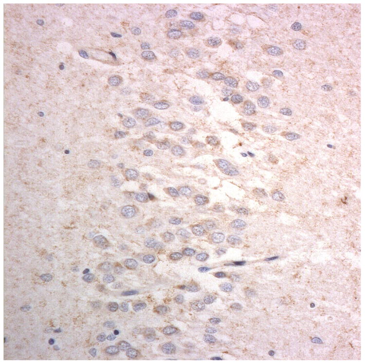
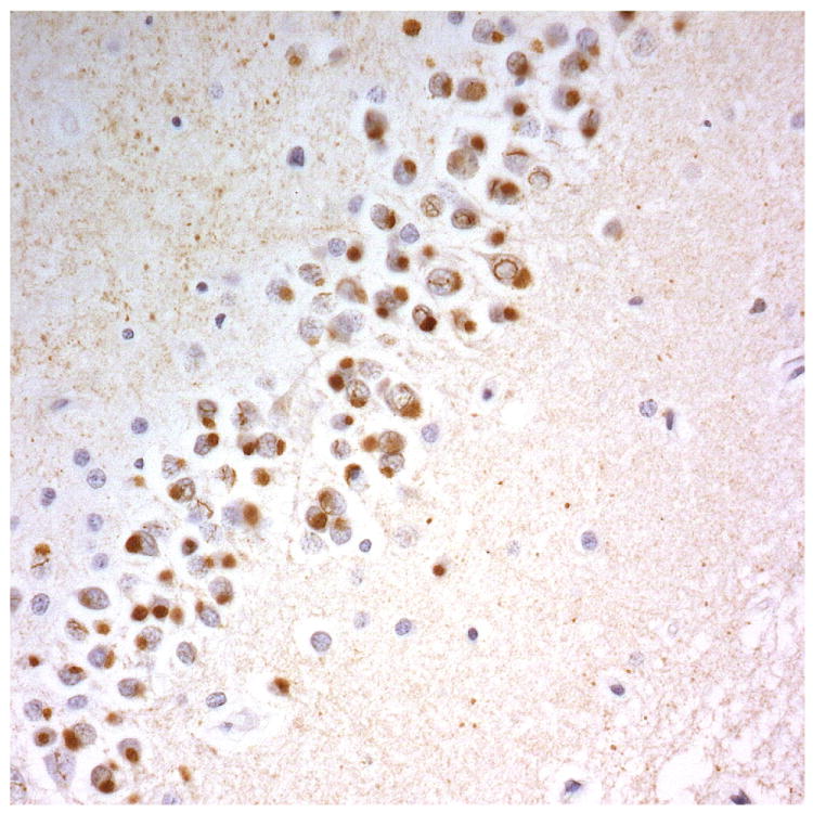
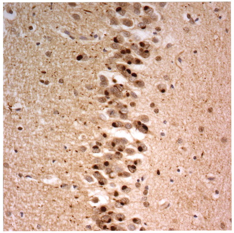
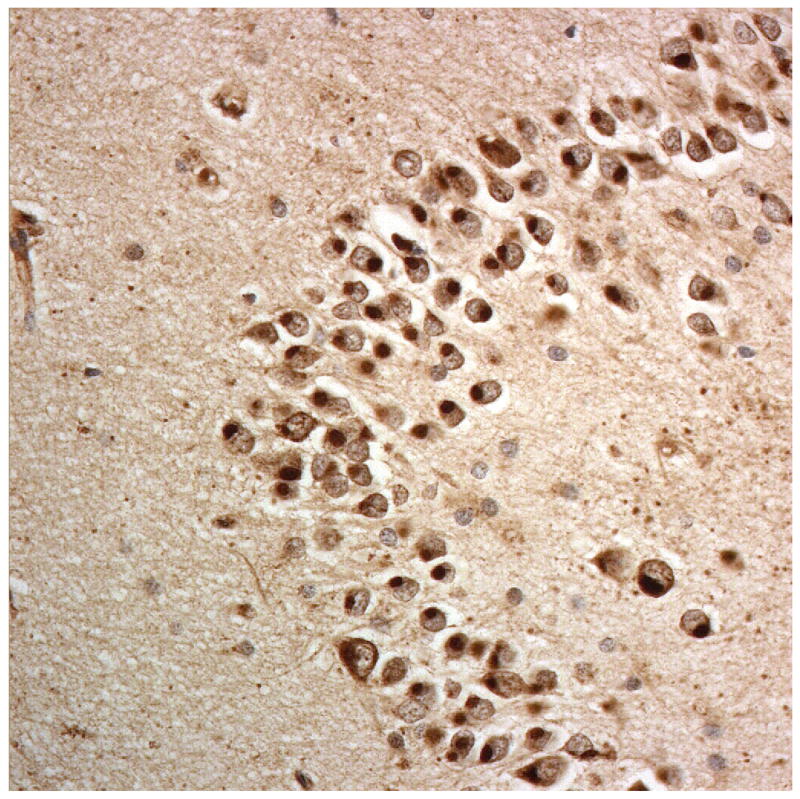
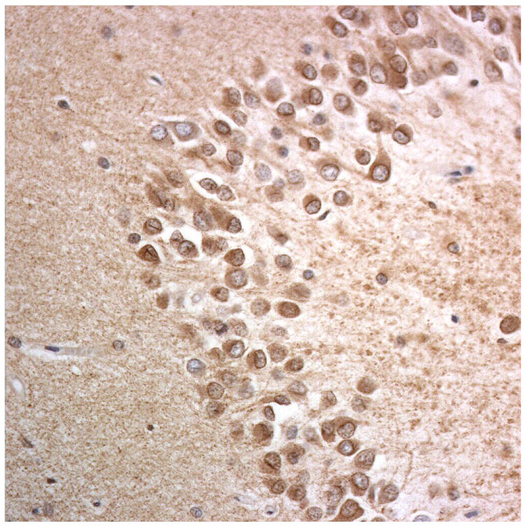
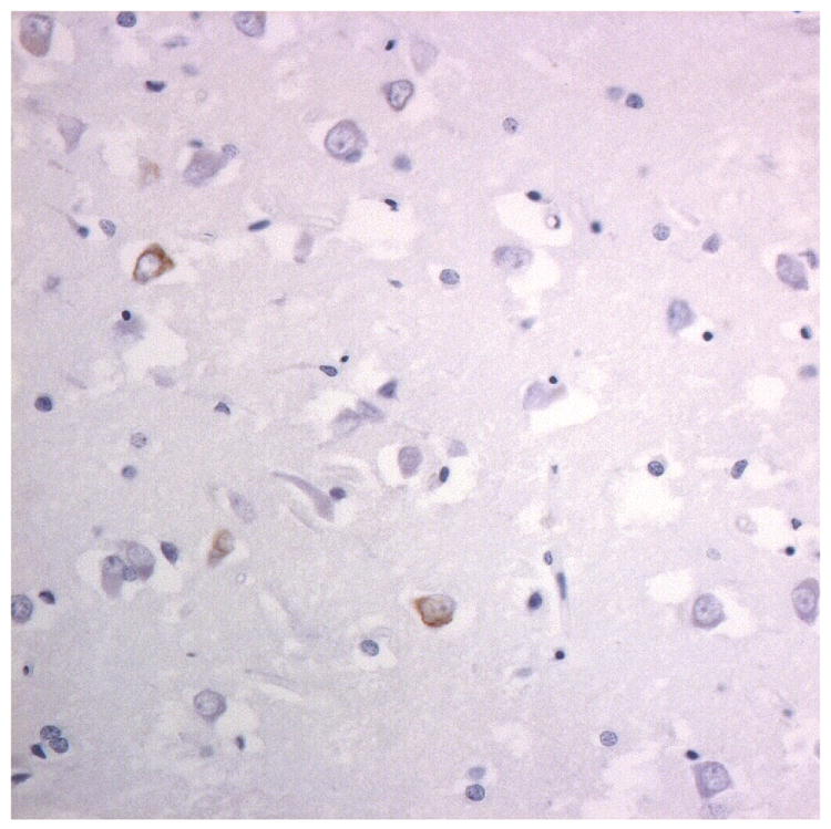
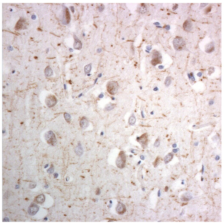
Results: The inclusion bodies in FTLD-FUS contained TRN1 and TAF15 and, to a lesser extent, EWS, but not TRN2. The patterns of immunostaining for TRN1 and TAF15 were very similar to that of FUS. None of these proteins was associated with tau or TDP-43 aggregations in FTLD.

Conclusions: Data suggest that FUS, TRN1 and TAF15 may participate in a functional pathway in an interdependent way, and imply that the function of TDP-43 may not necessarily be in parallel with, or complementary to, that of FUS, despite each protein sharing many similar structural elements.

Keywords: Ewing's sarcoma protein; TATA-binding protein-associated factor 15; TDP-43; frontotemporal lobar degeneration; fused in sarcoma; transportins.


Figures

Figure 1
Figure 1
Immunostaining for nuclear transport associated proteins in consecutive sections from aFTLD-U case #4. There is strong immunostaining of neuronal cytoplasmic inclusions (NCI) and intranuclear inclusions (NII) in dentate gyrus granule cells, in a similar distribution, for FUS (a), Abcam TRN1 (b) and TAF15 (c). The Sigma TRN1 antibody immunostains cell cytoplasm and nuclear membrane, but not NCI or NII (d). The EWS antibody only weakly immunostains a proportion of those NCI and NII immunolabelled by FUS, TRN1 and TAF15 (e). The TRN2 antibody immunolabels small amounts of granular material in the cytoplasm of both neurones and astrocytes (f). Immunoperoxidase – haematoxylin, all x40 microscope objective magnification.
Figure 1
Figure 1
Immunostaining for nuclear transport associated proteins in consecutive sections from aFTLD-U case #4. There is strong immunostaining of neuronal cytoplasmic inclusions (NCI) and intranuclear inclusions (NII) in dentate gyrus granule cells, in a similar distribution, for FUS (a), Abcam TRN1 (b) and TAF15 (c). The Sigma TRN1 antibody immunostains cell cytoplasm and nuclear membrane, but not NCI or NII (d). The EWS antibody only weakly immunostains a proportion of those NCI and NII immunolabelled by FUS, TRN1 and TAF15 (e). The TRN2 antibody immunolabels small amounts of granular material in the cytoplasm of both neurones and astrocytes (f). Immunoperoxidase – haematoxylin, all x40 microscope objective magnification.
Figure 2
Figure 2
Immunostaining for nuclear transport associated proteins in consecutive sections from NIFID case #6. There is strong immunostaining of neuronal cytoplasmic inclusions (NCI) and intranuclear inclusions (NII) in dentate gyrus granule cells, in a similar distribution, for FUS (a), Abcam TRN1 (b) and TAF15 (c). The Sigma TRN1 antibody immunostains cell cytoplasm and nuclear membrane, but not NCI or NII (d). In the temporal cortex (and dentate gyrus – not shown), the EWS antibody only weakly immunostains a small proportion of NCI immunolabelled by FUS, TRN1 and TAF15 (e). The TRN2 antibody immunolabels small amounts of granular material in the cytoplasm of both neurones and astrocytes (f). Immunoperoxidase – haematoxylin, all x40 microscope objective magnification.
Figure 2
Figure 2
Immunostaining for nuclear transport associated proteins in consecutive sections from NIFID case #6. There is strong immunostaining of neuronal cytoplasmic inclusions (NCI) and intranuclear inclusions (NII) in dentate gyrus granule cells, in a similar distribution, for FUS (a), Abcam TRN1 (b) and TAF15 (c). The Sigma TRN1 antibody immunostains cell cytoplasm and nuclear membrane, but not NCI or NII (d). In the temporal cortex (and dentate gyrus – not shown), the EWS antibody only weakly immunostains a small proportion of NCI immunolabelled by FUS, TRN1 and TAF15 (e). The TRN2 antibody immunolabels small amounts of granular material in the cytoplasm of both neurones and astrocytes (f). Immunoperoxidase – haematoxylin, all x40 microscope objective magnification.
Figure 3
Figure 3
Immunostaining for nuclear transport associated proteins in consecutive sections from BIBD case #8. There is strong immunostaining of neuronal cytoplasmic inclusions (NCI) in pyramidal cells of the motor cortex, in a similar distribution, for FUS (a), Abcam TRN1 (b) and TAF15 (c). The Sigma TRN1 antibody immunostains cell cytoplasm and nuclear membrane, but not NCI (d). The EWS antibody immunostains, less strongly, only a proportion of those NCI immunolabelled by FUS, TRN1 and TAF15 antibodies (e). The TRN2 antibody immunolabels small amounts of granular material in the cytoplasm of both neurones and astrocytes (f). Immunoperoxidase – haematoxylin, all x40 microscope objective magnification.
Figure 3
Figure 3
Immunostaining for nuclear transport associated proteins in consecutive sections from BIBD case #8. There is strong immunostaining of neuronal cytoplasmic inclusions (NCI) in pyramidal cells of the motor cortex, in a similar distribution, for FUS (a), Abcam TRN1 (b) and TAF15 (c). The Sigma TRN1 antibody immunostains cell cytoplasm and nuclear membrane, but not NCI (d). The EWS antibody immunostains, less strongly, only a proportion of those NCI immunolabelled by FUS, TRN1 and TAF15 antibodies (e). The TRN2 antibody immunolabels small amounts of granular material in the cytoplasm of both neurones and astrocytes (f). Immunoperoxidase – haematoxylin, all x40 microscope objective magnification.

Similar articles

Cited by

References

    1. Neary D, Snowden JS, Gustafson L, Passant U, Stuss D, Black S, Freedman M, Kertesz A, Robert PH, Albert M, Boone K, Miller BL, Cummings J, Benson DF. Frontotemporal lobar degeneration: a consensus on clinical diagnostic criteria. Neurology. 1998;51:1546–54. - PubMed
    1. Neary D, Snowden JS, Mann DMA, Northen B, Goulding PJ, MacDermott N. Frontal lobe dementia and motor neurone disease. J Neurol Neurosurg Psychiatry. 1990;53:23–32. - PMC - PubMed
    1. Strong MJ, Grace GM, Freedman M, Lomen-Hoerth C, Woolley S, Goldstein LH, Murphy J, Shoesmith C, Rosenfeld J, Leigh PN, Bruijn L, Ince P, Figlewicz D. Consensus criteria for the diagnosis of frontotemporal cognitive and behavioural syndromes in amyotrophic lateral sclerosis. Amyotroph Lateral Scler. 2009;10:131–46. - PubMed
    1. Cairns NJ, Bigio EH, Mackenzie IRA, Neumann M, Lee VMY, Hatanpaa KJ, White CL, III, Schneider JA, Tenenholz Grinberg L, Halliday G, Duyckaerts C, Lowe JS, Holm IE, Tolnay M, Okamoto K, Yokoo H, Murayama S, Woulfe J, Munoz DG, Dickson DW, Ince PG, Trojanowski JQ, Mann DMA. Neuropathologic diagnostic and nosologic criteria for frontotemporal lobar degeneration: consensus of the Consortium for Frontotemporal Lobar Degeneration. Acta Neuropathol. 2007;114:2–22. - PMC - PubMed
    1. Arai T, Hasegawa M, Akiyama H, Ikeda K, Nonaka T, Mori H, Mann D, Tsuchiya K, Yoshida M, Hashizume Y, Oda T. TDP-43 is a component of ubiquitin-positive tau-negative inclusions in frontotemporal lobar degeneration and amyotrophic lateral sclerosis. Biochem Biophys Res Commun. 2006;351:602–11. - PubMed

Publication types

LinkOut - more resources