. 2012 Apr 12:12:146.
doi: 10.1186/1471-2407-12-146.

HOXA1 is overexpressed in oral squamous cell carcinomas and its expression is correlated with poor prognosis

Affiliations

HOXA1 is overexpressed in oral squamous cell carcinomas and its expression is correlated with poor prognosis

Carolina Cavalcante Bitu et al. BMC Cancer. .

Abstract

Background: HOX genes encode homeodomain-containing transcription factors involved in the regulation of cellular proliferation and differentiation during embryogenesis. However, members of this family demonstrated oncogenic properties in some malignancies. The present study investigated whether genes of the HOXA cluster play a role in oral cancer.

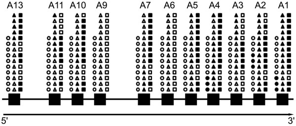
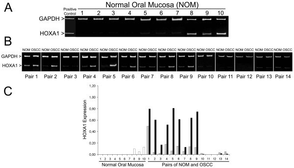
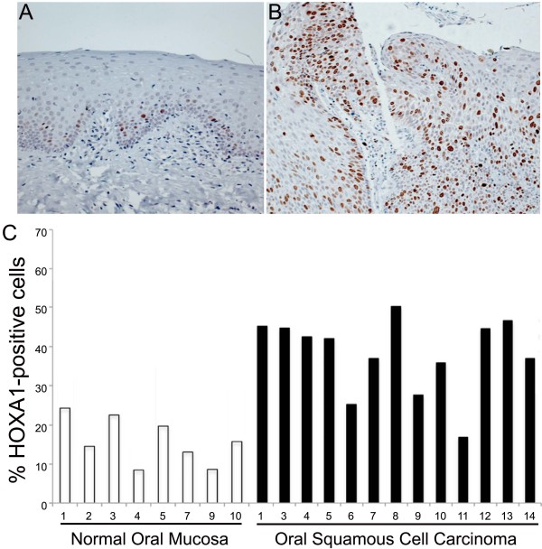
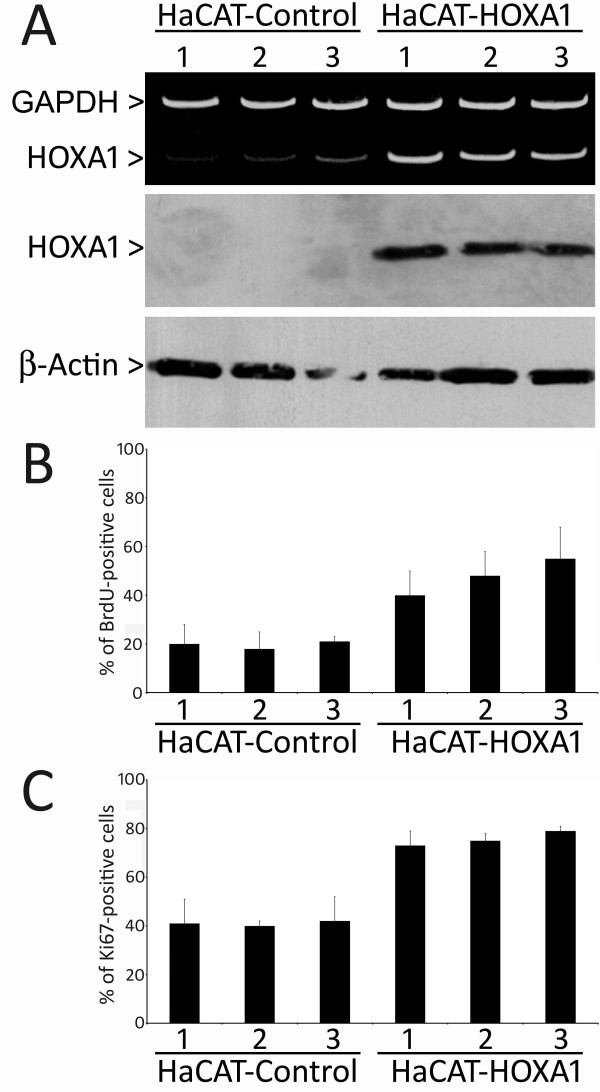
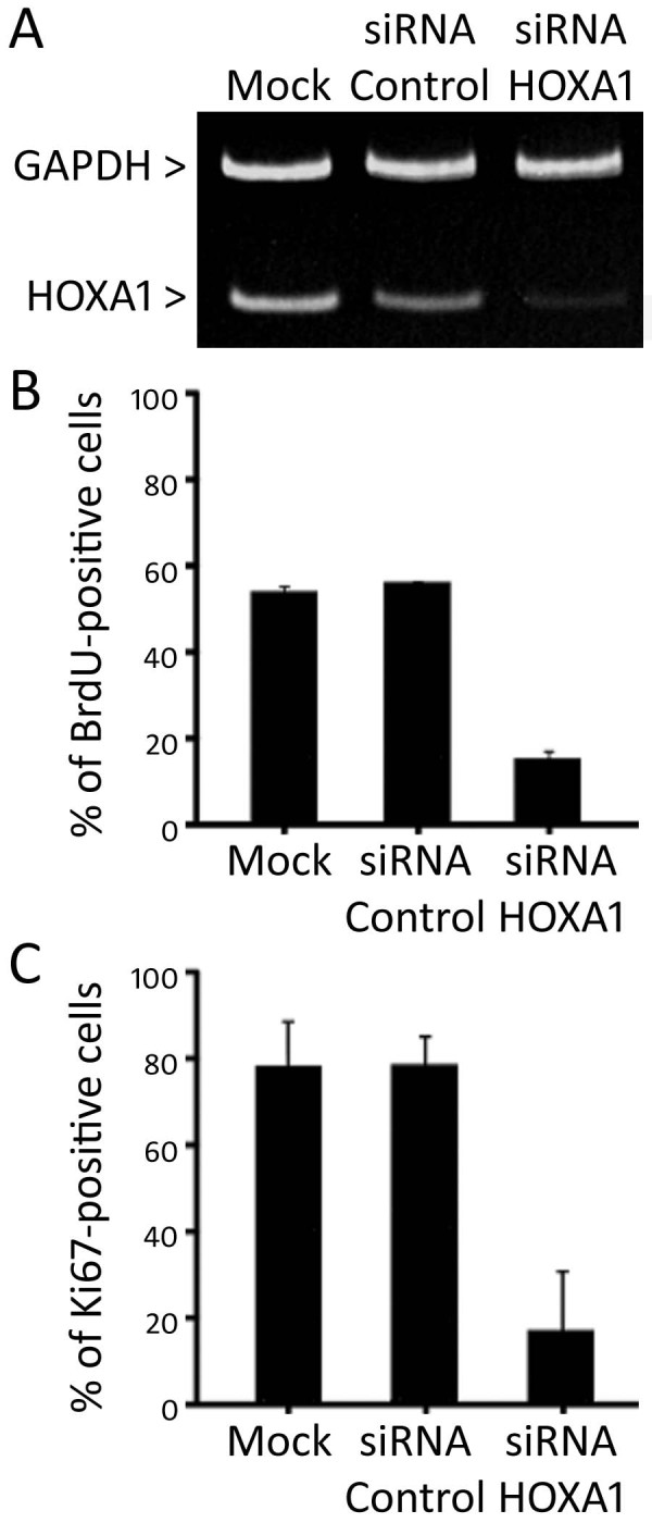
Methods: In order to identify differentially expressed HOXA genes, duplex RT-PCR in oral samples from healthy mucosa and squamous cell carcinoma was used. The effects of HOXA1 on proliferation, apoptosis, adhesion, invasion, epithelial-mesenchymal transition (EMT) and anchorage-independent growth were assessed in cells with up- and down-regulation of HOXA1. Immunohistochemical analysis using a tissue microarray (TMA) containing 127 oral squamous cell carcinomas (OSCC) was performed to determine the prognostic role of HOXA1 expression.

Results: We showed that transcripts of HOXA genes are more abundant in OSCC than in healthy oral mucosa. In particular, HOXA1, which has been described as one of the HOX members that plays an important role in tumorigenesis, was significantly more expressed in OSCCs compared to healthy oral mucosas. Further analysis demonstrated that overexpression of HOXA1 in HaCAT human epithelial cells promotes proliferation, whereas downregulation of HOXA1 in human OSCC cells (SCC9 cells) decreases it. Enforced HOXA1 expression in HaCAT cells was not capable of modulating other events related to tumorigenesis, including apoptosis, adhesion, invasion, EMT and anchorage-independent growth. A high number of HOXA1-positive cells was significantly associated with T stage, N stage, tumor differentiation and proliferative potential of the tumors, and was predictive of poor survival. In multivariate analysis, HOXA1 was an independent prognostic factor for OSCC patients (HR: 2.68; 95% CI: 1.59-2.97; p = 0.026).

Conclusion: Our findings indicate that HOXA1 may contribute to oral carcinogenesis by increasing tumor cell proliferation, and suggest that HOXA1 expression might be helpful as a prognostic marker for patients with OSCC.


Figures

Figure 1
Figure 1
Diagram of expression of the members of HOXA locus in samples from healthy oral mucosa, normal-looking oral mucosa adjacent to OSCC, and OSCC. Each symbol represents one specific sample, and their positions are preserved throughout the image. Circles represent healthy oral mucosa derived from patients without contact with the main oral cancer risk factors, and triangles and squares represent normal-looking mucosa and OSCC from the same patient, respectively. Open symbols indicate silent (inactive) HOXA genes, whereas closed symbols indicate active genes. Note the abundance of active genes in OSCC samples.
Figure 2
Figure 2
HOXA1 is overexpressed in OSCCs. (A) Analysis of HOXA1 and GAPDH by duplex RT-PCR on samples of healthy oral mucosa from patients without contact with the main risk factors for oral cancer and (B) on pairs of OSCC and adjacent normal-looking oral mucosa from the same patient. (C) Densitometric analysis of the HOXA1 bands demonstrated a significant higher expression in OSCCs compared to healthy oral mucosas from patients without recognized OSCC-risk factors (p < 0.001).
Figure 3
Figure 3
Immunohistochemical detection of HOXA1 in healthy oral mucosa and OSCC. Representative samples of healthy oral mucosa (A) and OSCC (B) of this study. In the healthy mucosa, HOXA1 was clearly limited to the nucleus of the epithelial cells located in the basal and suprabasal layers, whereas in the OSCCs, HOXA1-positive cells were broadly observed in the tumor (Original magnification x200). (C) The percentage of HOXA1-positive cells was significantly higher in OSCCs than in healthy oral mucosas (p < 0.001).
Figure 4
Figure 4
Overexpression of HOXA1 induces cellular proliferation. (A) Representative duplex RT-PCR and western blot analysis of HOXA1 in HaCAT-Control and HaCAT-HOXA1 transfectants, revealing an increase in HOXA1 levels in overexpressing clones. Assays measuring BrdU incorporation (B) and Ki67 expression (C) demonstrated that HOXA1 overexpressing cell lines have a statistically significant increase in proliferation as compared to control cells (for BrdU index p < 0.01 between groups, and for Ki67 index p < 0.05 between groups). The labeling index of BrdU and Ki67 corresponds to the mean percentage of positive cells of 3 experiments for each cell line.
Figure 5
Figure 5
Inhibition of HOXA1 by siRNA in SCC9 cells decreases cellular proliferation. (A) siRNA working against HOXA1 significantly decreased HOXA1 levels as revealed by representative duplex RT-PCR. (B) BrdU incorporation and Ki67 immunocytochemical expression (C) assays showed a statistically significant decrease in proliferation when HOXA1 was downregulated via siRNA (for BrdU index p < 0.01 and for Ki67 index p < 0.05). Data correspond to the mean percentage of positive cells of 3 experiments.
Figure 6
Figure 6
Expression of HOXA1 is associated with shortened overall survival of patients with OSCC. (A) The overall survival analysis according to the Kaplan-Meier method for HOXA1 immunoexpression revealed that high HOXA1 expression is associated with poor prognosis with a 5-year survival of 55.5% (p = 0.0073). (B) Disease-specific overall survival for patients with high expression of HOXA1 was even shorter (p = 0.0048). (C) Five-year disease-free survival was not associated with HOXA1 immunoexpression in OSCC tumors (p = 0.27).

References

    1. Scully C, Bagan J. Oral squamous cell carcinoma: overview of current understanding of aetiopathogenesis and clinical implications. Oral Dis. 2009;15(6):388–399. doi: 10.1111/j.1601-0825.2009.01563.x. - DOI - PubMed
    1. Warnakulasuriya S. Global epidemiology of oral and oropharyngeal cancer. Oral Oncol. 2009;45(4-5):309–316. doi: 10.1016/j.oraloncology.2008.06.002. - DOI - PubMed
    1. Hassan NM, Hamada J, Murai T, Seino A, Takahas'hi Y, Tada M, Zhang X, Kashiwazaki H, Yamazaki Y, Inoue N, Moriuchi T. Aberrant expression of HOX genes in oral dysplasia and squamous cell carcinoma tissues. Oncol Res. 2006;16(5):217–224. - PubMed
    1. Grier DG, Thompson A, Kwasniewska A, McGonigle GJ, Halliday HL, Lappin TR. The pathophysiology of HOX genes and their role in cancer. J Pathol. 2005;205(2):154–171. doi: 10.1002/path.1710. - DOI - PubMed
    1. Abate-Shen C. Deregulated homeobox gene expression in cancer: cause or consequence? Nat Rev Cancer. 2002;2(10):777–785. doi: 10.1038/nrc907. - DOI - PubMed

Publication types

MeSH terms