. 2012 Jul;33(20):5124-9.
doi: 10.1016/j.biomaterials.2012.03.066. Epub 2012 Apr 11.

Hollow silica and silica-boron nano/microparticles for contrast-enhanced ultrasound to detect small tumors

Affiliations

Hollow silica and silica-boron nano/microparticles for contrast-enhanced ultrasound to detect small tumors

Alexander Liberman et al. Biomaterials. 2012 Jul.

Abstract

Diagnosing tumors at an early stage when they are easily curable and may not require systemic chemotherapy remains a challenge to clinicians. In order to improve early cancer detection, gas filled hollow boron-doped silica particles have been developed, which can be used for ultrasound-guided breast conservation therapy. The particles are synthesized using a polystyrene template and subsequently calcinated to create hollow, rigid nanoporous microspheres. The microshells are filled with perfluoropentane vapor. Studies were performed in phantoms to optimize particle concentration, injection dose, and the ultrasound settings such as pulse frequency and mechanical index. In vitro studies have shown that these particles can be continuously imaged by US up to 48 min and their signal lifetime persisted for 5 days. These particles could potentially be given by intravenous injection and, in conjunction with contrast-enhanced ultrasound, be utilized as a screening tool to detect smaller breast cancers before they are detectible by traditional mammography.


Figures

Fig. 1
Fig. 1
SEM images of porous hollow silica nano and microshells prepared using a templating sol-gel process. (A) 100 nm diameter nanoshells; (B) 500 nm nanoshells; (C) 2 μm microshells. Note: all have a shell thickness of ~10 nm and a uniform size distribution.
Fig. 2
Fig. 2
Continuous imaging time in a chicken breast phantom. A 50 μl bolus of 2 mg/ml of 2 μm sized gas filled nanoshells were injected into chicken breast tissue and imaged continuously under color Doppler with different mechanical indices until no usable signal was detectable. The continuous imaging time was greater at low mechanical index.
Fig. 3
Fig. 3
Persistence in a phantom. (A) Color Doppler image (CDI) of 50 μl of 2 mg/ml 2 μm gas filled microshells after injection into a chicken breast phantom kept at 25 °C. (B) CDI 96 h after injection. (C) CDI signal area vs. time. As seen here, there was substantial signal present from the same injection up to 120 h after initial injection which was consistent with high gas retention, high in vitro stability, and low color Doppler signal decay.
Fig. 4
Fig. 4
Area of CDI signal vs. transducer frequency. A 50 μl bolus of 2 mg/ml 2 μm of gas filled microshells were injected into a chicken breast phantom and imaged at multiple frequencies using a 15L8 Sequoia transducer. As frequency decreased, signal area increased exponentially. The red dots are the raw data points and the black line is the fitted equation. f(x) = 0.74 e−0.38x +4.13 × 10¹⁴e−4.9x r2 > 0:99. (For interpretation of the references to colour in this figure legend, the reader is referred to the web version of this article.)
Fig. 5
Fig. 5
Effect of ascending mechanical index (MI) on CPS imaging of gas filled silica nano and Microshells. Particles were imaged using CPS mode with MI scaling from 0.06 to 1.9. (A) CPS image of 100 μg/ml of gas filled 2 μm microparticles at MI = 0.87. Each gold speck is a single event (white arrows). (B) CPS image 100 μg/ml of gas filled 2 μm microparticles at MI = 1.4. The large density of gold specks corresponds to a large number of particles being imaged. (C) Signal brightness plotted against MI for 2 μm gas filled microshells. (D) Signal brightness vs. MI for gas filled 500 nm silica nanoshells. Note that the 500 nm particles generated less signal than the 2 μm particles at the same MI.
Fig. 6
Fig. 6
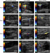
Persistence in an in vivo model. 50 μl of control microbubbles, 2 μm shells and 500 nm shells were injected into New Zealand White Rabbit thighs and imaged over the course of four days. Shown in the left columns are the control microbubbles; 50 μl were injected containing 108 microbubbles/ml. All injections were imaged at an MI of 1.9 at 7 MHz with color Doppler using the Siemens Sequoia. Day 0 corresponds to imaging within 15 min of the injection. Note that signal persisted for 4 days when either formulation of silica particles were injected. Microbubbles given as 108 (left column) or 1010 (not shown) could not be detected 1 day after injection.
Fig. 7
Fig. 7
Testing of gas filled silica microshells in a Mouse. (A) Dissected nu/nu mouse with an interperitoneal IGROV-1 ovarian tumor (red arrow white mass on right side of image). 200 μg of 2 μm silica shells diluted into 3 ml of saline and injected into the peritoneum and then perfused into the blood. (B) CPS imaging of the particles through a cross section of the tumor 1 h after quasi IV injection. (C) B-mode image through a cross section of the tumor 1 h after quasi IV injection. (D) Overlay image using several frames from CPS imaging and B-mode to show an integrated heat map of signal from the particles. For all the images, the red arrow points to the tumor, the green arrow points to the spinal column and the blue arrow points to the bottom of the mouse. (For interpretation of the references to colour in this figure legend, the reader is referred to the web version of this article.)

References

    1. Klimberg VS, Harms S, Korourian S. Assessing margin status. Surg Oncol. 1999;(2):77–84. - PubMed
    1. Mullenix PS, Cuadrado DG, Steele SR, Martin MJ, See CS, Beitler AL, et al. Secondary operations are frequently required to complete the surgical phase of therapy in the era of breast conservation and sentinel lymph node biopsy. Am J Surg. 2004;187(5):643–6. - PubMed
    1. Singletary SE. Surgical margins in patients with early-stage breast cancer treated with breast conservation therapy. Am J Surg. 2002;184(5):383–93. - PubMed
    1. Gray RJ, Salud C, Nguyen K, Dauway E, Friedland J, Berman C, et al. Randomized prospective evaluation of a novel technique for biopsy or lumpectomy of nonpalpable breast lesions: radioactive seed versus wire localization. Ann Surg Oncol. 2011;8(9):711–5. - PubMed
    1. Gray RJ, Pockaj BA, Karstaed PJ, C.Roarke M. Radioactive seed localization of nonpalpable breast lesions is better than wire localization. Am J Surg. 2004;188(4):377–80. - PubMed

Publication types