. 2012 Aug;17(7):921-32.
doi: 10.1177/1087057112442960. Epub 2012 Apr 12.

Evaluation of a luciferase-based reporter assay as a screen for inhibitors of estrogen-ERα-induced proliferation of breast cancer cells

Affiliations

Evaluation of a luciferase-based reporter assay as a screen for inhibitors of estrogen-ERα-induced proliferation of breast cancer cells

Neal Andruska et al. J Biomol Screen. 2012 Aug.

Abstract

Estrogens, acting through estrogen receptor α (ERα), stimulate breast cancer proliferation, making ERα an attractive drug target. Since 384-well format screens for inhibitors of proliferation can be challenging for some cells, inhibition of luciferase-based reporters is often used as a surrogate end point. To identify novel small-molecule inhibitors of 17β-estradiol (E(2))-ERα-stimulated cell proliferation, we established a cell-based screen for inhibitors of E(2)-ERα induction of an estrogen response element (ERE)(3)-luciferase reporter. Seventy-five "hits" were evaluated in tiered follow-up assays to identify where hits failed to progress and evaluate their effectiveness as inhibitors of E(2)-ERα-induced proliferation of breast cancer cells. Only 8 of 75 hits from the luciferase screen inhibited estrogen-induced proliferation of ERα-positive MCF-7 and T47D cells but not control ERα-negative MDA-MB-231 cells. Although 12% of compounds inhibited E(2)-ERα-stimulated proliferation in only one of the ERα-positive cell lines, 40% of compounds were toxic and inhibited growth of all the cell lines, and ~37% exhibited little or no ability to inhibit E(2)-ERα-stimulated cell proliferation. Representative compounds were evaluated in more detail, and a lead ERα inhibitor was identified.


Figures

FIG. 1
FIG. 1
The (ERE)3-luciferase based assay. (A) Dose response study of E2-ERα induction of (ERE)3-luciferase. The data represents the average ± S.E.M. of quadruplicate assays carried out in 96 well plates. (B) Assessment of screen robustness using Z’-factor.
FIG. 2
FIG. 2
Primary screening data for the 75 representative compounds selected for further characterization. For each compound, percent inhibition of DHT-AR-stimulated ARE-luciferase activity (black bars) and E2-ERα-stimulated (ERE)3-luciferase activity (grey bars) is shown. SSMD scores for small molecules are shown in Supplemental Figure 2.
FIG 3
FIG 3
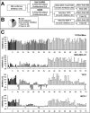
Summary of assays used to evaluate inhibitors of ERα-mediated gene expression as a surrogate marker for inhibitors of E2-dependent growth. (A) Flow chart showing the breakdown by category of compounds screened for reporter activity and growth inhibition. (B) Summary of the classes of compounds identified. (C) Compounds were further assessed for (from top to bottom) their ability to inhibit (ERE)3-luciferase activity (T47D-KBluc), growth of ERα negative MDA-MB-231 breast cancer cells (MDAMB-231), E2-ERα-dependent proliferation of ERα positive T47D, and MCF-7 breast cancer cells. Mean inhibition represents the average of three-independent experiments ± S. E.M. Numerical data is shown in Supplemental Table 1.
FIG. 4
FIG. 4
Characterization of representative inhibitors. (A) Structures of small molecules selected to represent each class of compounds for further evaluation. (B) Dose-response curves for inhibition of (ERE)3-luciferase by selected compounds.T47D-KBluc cells were maintained for 3 days in 10% CD-FBS and then incubated for 24 hours in the presence of 10 nM E2 and the indicated concentrations of the test compounds. Cells were harvested and assayed for luciferase activity. Mean percent inhibition represents the average of three-independent experiments ± S. E.M.
FIG 5
FIG 5
Dose-response studies of the effects of the inhibitors on cell proliferation. Dose response studies were used to evaluate the effect on the E2-ERα-dependent proliferation of MCF-7 and T47D calls and on the growth of ERα negative MDA-MB-231 for (A) Compound 4, (B) Compound 45, (C) Compound 14, and (D) Compound 11. Cells received the indicated concentrations of each inhibitor in DMSO. After 3.5 days cell number was quantified by MTS. ERα positive cell lines were also treated with 1 μM ICI 182,780, a known ER antagonist. Mean percent growth inhibition represents the average of three-independent experiments ± S. E.M.
FIG 6
FIG 6
Effect of the 3 inhibitors on ERα levels and on the induction of pS2 mRNA. The effects of the three compounds on the level of ERα was evaluated in Western blots of extracts from (A) T47D cells and (B) MCF-7 cells. ICI 182,780 (ICI) and TPSF, known down-regulators of ERα were used as controls. Cells were treated for 24 hours with or without 10 nM E2, or a combination of 10 nM E2 and 1 μM ICI, 10 μM TPSF, 5 μM Compound 4, 10 μM Compound 11, or 10 μM Compound 14. (C) Compounds were tested for their ability to inhibit E2-ERα induction of pS2 mRNA. pS2 mRNA levels were quantified by qRT-PCR, following 24-hour treatment of T47D-KBluc cells with and without 10 nM E2, or a combination of 10 nM E2 and each compound at the concentrations listed above. Average fold-change represents the average of three-independent experiments ± S. E.M.

References

    1. Fan F, Wood KV. Bioluminescent assays for high-throughput screening. Assay Drug Dev. Technol. 2007;5:127–36. - PubMed
    1. Auld DS, Thorne N, Maguire WF, Inglese J. Mechanism of PTC124 activity in cell-based luciferase assays of nonsense codon suppression. Proc. Natl. Acad. Sci. U.S.A. 2009;106:3585–3590. - PMC - PubMed
    1. Inglese J, Johnson RL, Simeonov A, Xia M, Zheng W, Austin CP, Auld DS. High-throughput screening assays for the identification of chemical probes. Nat. Chem. Biol. 2007;3:466–479. - PubMed
    1. Lewis JS, Cheng D, Jordan VC. Targeting oestrogen to kill the cancer but not the patient. Br. J. Cancer. 2004;90:944–949. - PMC - PubMed
    1. Jordan VC. Chemoprevention with antiestrogens: the beginning of the end for breast cancer. Daniel G. Miller Lecture. Ann. N.Y. Acad. Sci. 2001;952:60–72. - PubMed

Publication types