Review
. 2012;88(4):129-43.
doi: 10.2183/pjab.88.129.

A world of sphingolipids and glycolipids in the brain--novel functions of simple lipids modified with glucose

Affiliations
Review

A world of sphingolipids and glycolipids in the brain--novel functions of simple lipids modified with glucose

Yoshio Hirabayashi. Proc Jpn Acad Ser B Phys Biol Sci. 2012.

Abstract

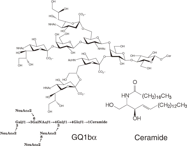
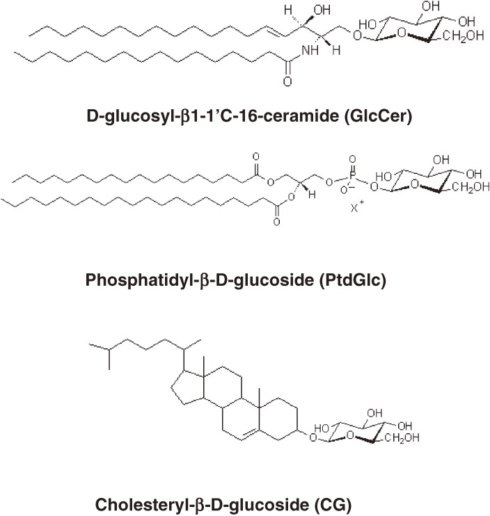
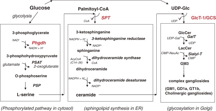
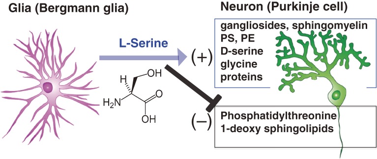
Glycosphingolipids (GSLs) are present on cell surface membranes and are particularly abundant in the brain. Since over 300-400 GSLs are synthesized from glucosylceramide (GlcCer), GlcCer is believed to only serve as the source of most GSLs, including sialic acid-containing GSLs or gangliosides, in the brain. Recent studies, however, suggest that GlcCer itself plays a role in the heat stress response, as it functions as a glucose donor for the synthesis of cholesterylglucoside, a lipid mediator in heat stress responses in animals. GlcCer in adipose tissues is also thought to be involved in mechanisms that regulate energy (sugar and lipid) metabolism. Our extensive structural study revealed an additional novel glucosylated membrane lipid, called phosphatidylglucoside, in developing rodent brains and human neutrophils. These lipids, all modified with glucose, are enriched in lipid rafts and play important roles in basic cellular processes. Here, I summarize the recent progress regarding these glucosylated lipids and their biosynthesis and regulation in the central nervous system (CNS).


Figures

Figure 1.
Figure 1.
Cholinergic gangliosides are one the most complex type of gangliosides in the CNS. Q1bα, which was first isolated from bovine brain,5) is expressed in cholinergic neurons. Recent studies indicate that Q1bα may play a role in pathophysiological processes leading to Alzheimer’s disease.92)
Figure 2.
Figure 2.
Three lipids modified with glucose present in mammalian membranes.
Figure 3.
Figure 3.
Sphingolipids and sphingoglycolipids are synthesized from glucose, amino acid, and fatty acid. The ceramide molecule contains a nitrogen atom that is derived from the amino acid L-serine. To synthesize the precursor lipid for GSLs, glucosylceramide (GlcCer), glucose (UDP-Glc), L-serine, and palmitoyl-CoA are required. Thus, it is understandable that GlcCer and its related compounds are involved in the regulation of energy metabolism and in the pathophysiology of diabetes mellitus.
Figure 4.
Figure 4.
Metabolic relationship between neurons and glia in cerebellum mediated through L-serine. Phgdh, a key enzyme for L-serine synthesis, is upregulated in Bergmann glia cells but downregulated in Purkinje neurons. Thus, Purkinje neurons are dependent on glial L-serine. L-serine stimulates the synthesis of membrane lipids such as sphingolipids. Importantly, L-serine inhibits the synthesis of phosphatidylthreonine and toxic lipids such as 1-deoxsphinganine.
Figure 5.
Figure 5.
Occurrence of unusual sphingolipids: 1-deoxysphinganine and 1-desoxymethylsphinganine
Figure 6.
Figure 6.
Developmental changes in PtdGlc expression in mouse cerebral cortex. In all images, cells labeled with DIM21 are green. Brain lipid-binding protein (BLBP) is a radial glia marker. DIM21-positive cells express the astrocyte marker GFAP (arrowheads) at E18.5.83)
Figure 7.
Figure 7.
Exogenous addition of lyso-phosphatidylglucoside induces growth cone collapse of rat DRG neurons. DRG neurons were isolated from an E12.5 rat. Actin filaments were visualized with rhodamine-labeled phalloidin (M. Yamazaki et al., unpublished data). A, Control; B, LPA (100 µM); C, lyso-PtdGlc (10 µM). The lyso form of PtGlc showed more potent activity for growth cone collapse.

References

    1. Smith W.L., Merrill A.H., Jr. (2002) Sphingolipid metabolism and signaling minireview series. J. Biol. Chem. 277 (29), 25841–25842 - PubMed
    1. Ando S., Tanaka Y., Kobayashi S., Fukui F., Iwamoto M., Waki H., Tai T., Hirabayashi Y. (2004) Synaptic function of cholinergic-specific Chol-1α ganglioside. Neurochem. Res. 29 (4), 857–867 - PubMed
    1. Furuya S., Irie F., Hashikawa T., Nakazawa K., Kozakai A., Hasegawa A., Sudo K., Hirabayashi Y. (1994) Ganglioside GD1α in cerebellar Purkinje cells. Its specific absence in mouse mutants with Purkinje cell abnormality and altered immunoreactivity in response to conjunctive stimuli causing longterm desensitization. J. Biol. Chem. 269 (51), 32418–32425 - PubMed
    1. Hidari K.I., Irie F., Suzuki M., Kon K., Ando S., Hirabayashi Y. (1993) A novel ganglioside with a free amino group in bovine brain. Biochem. J. 296 (Pt 1), 259–263 - PMC - PubMed
    1. Hirabayashi Y., Nakao T., Irie F., Whittaker V.P., Kon K., Ando S. (1992) Structural characterization of a novel cholinergic neuron-specific ganglioside in bovine brain. J. Biol. Chem. 267 (18), 12973–12978 - PubMed