. 2012 May 8;109(19):7439-44.
doi: 10.1073/pnas.1200566109. Epub 2012 Apr 12.

Identification of human neutralizing antibodies that bind to complex epitopes on dengue virions

Affiliations

Identification of human neutralizing antibodies that bind to complex epitopes on dengue virions

Ruklanthi de Alwis et al. Proc Natl Acad Sci U S A. .

Abstract

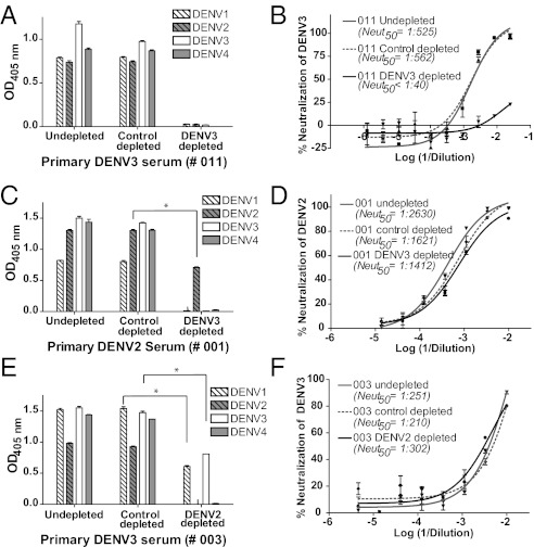
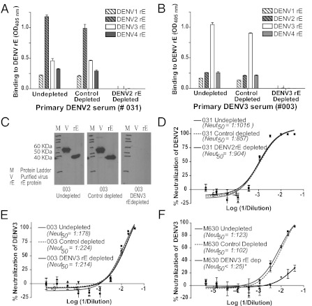
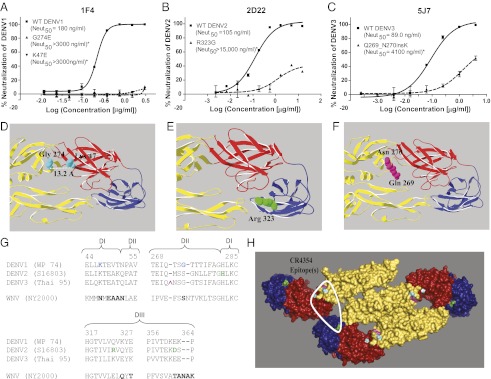
Dengue is a mosquito-borne flavivirus that is spreading at an unprecedented rate and has developed into a major health and economic burden in over 50 countries. Even though infected individuals develop potent and long-lasting serotype-specific neutralizing antibodies (Abs), the epitopes engaged by human neutralizing Abs have not been identified. Here, we demonstrate that the dengue virus (DENV)-specific serum Ab response in humans consists of a large fraction of cross-reactive, poorly neutralizing Abs and a small fraction of serotype-specific, potently inhibitory Abs. Although many mouse-generated, strongly neutralizing monoclonal antibodies (mAbs) recognize epitopes that are present on recombinant DENV envelope (E) proteins, unexpectedly, the majority of neutralizing Abs in human immune sera bound to intact virions but not to the ectodomain of purified soluble E proteins. These conclusions with polyclonal Abs were confirmed with newly generated human mAbs derived from DENV-immune individuals. Two of three strongly neutralizing human mAbs bound to E protein epitopes that were preserved on the virion but not on recombinant E (rE) protein. We propose that humans produce Abs that neutralize DENV infection by binding a complex, quaternary structure epitope that is expressed only when E proteins are assembled on a virus particle. Mapping studies indicate that this epitope has a footprint that spans adjacent E protein dimers and includes residues at the hinge between domains I and II of E protein. These results have significant implications for the DENV Ab and vaccine field.


Conflict of interest statement

The authors declare no conflict of interest.

Figures

Fig. 1.
Fig. 1.
Binding and neutralization properties of primary DENV-immune sera depleted of total or cross-reactive DENV-binding Abs. Total DENV-specific Abs were removed from DENV3 primary immune serum (e.g., subject 011) using polystyrene beads coated with purified DENV3 and tested for DENV binding (A) and neutralization (B). The sera depleted with the homologous serotype did not bind to any of the four DENVs and failed to neutralize DENV3. Similar results were observed for four other primary immune sera (2 DENV2 and 2 DENV3 sera) depleted with the homologous serotype responsible for infection. Primary DENV2 (C and D) and DENV3 (E and F) immune sera were depleted of cross-reactive Abs using beads coated with virus of a heterologous serotype and tested for DENV binding (C and E) and neutralization of the homologous serotype (D and F). Immune sera depleted of cross-reactive Abs contained type-specific Abs that bound to virus from the homologous serotype only. Immune sera depleted of cross-reactive Abs were as potently neutralizing as undepleted or control-depleted sera. Results presented here for cross-reactive Ab depletions are representative of data obtained with four primary DENV2 and three primary DENV3 human immune sera (Table 1). *P < 0.001 by an unpaired Student t test of mean binding values.
Fig. 2.
Fig. 2.
Binding and neutralization properties of primary DENV-immune sera depleted of rE-binding Abs. DENV rE from the homotypic strain was coupled covalently to agarose beads and incubated with the relevant DENV-immune sera to deplete DENV rE-specific Abs. (A and B) Binding of immune sera to rE protein. Primary DENV2 (A) and DENV3-immune (B) sera were depleted with DENV2 and DENV3 rE proteins, respectively, and binding to rE protein from each of the four serotypes was measured by ELISA. Depletion with the rE from the homologous serotype led to a loss of binding to rE protein from each of the four serotypes. (C) Successful removal of all rE-reactive Abs from sera (e.g., primary DENV3-immune subject 003) also was confirmed by Western blot analysis. Purified homotypic DENV (700 ng per well) and rE protein (500 ng per well) were electrophoresed, transferred to nitrocellulose membrane, and probed with undepleted, control-depleted, or rE-depleted sera (at a 1:1,000 dilution). (D and E) Neutralization of the homologous DENV by rE-depleted sera was measured using a U937 + DC-SIGN flow cytometry-based assay. Homologous DENV neutralization by primary DENV2 (D; subject 031) and primary DENV3 (E; subject 003) human immune sera depleted of rE-binding Abs was tested. No reduction in neutralization potency was observed following removal of rE-binding Abs from either of these two serum samples. A total of six primary immune sera were depleted of rE-binding Abs and tested (Table 2). (F) Nonhuman primates vaccinated with rE develop neutralizing Abs that can be depleted with rE antigen. Rhesus macaques (M. mulatta) were vaccinated and boosted with an α-virus vector expressing DENV3 E ectodomain, and sera were collected 10 wk postvaccination. Depletion of rE-binding Abs from sera of vaccinated animals (e.g., M630) removed greater than 98% (value estimated by comparing Neut50 values between control-depleted and rE-depleted sera) of the neutralizing Abs. Data are representative of two vaccinated rhesus macaque controls.
Fig. 3.
Fig. 3.
Epitope mapping of escape mutants generated from type-specific neutralizing hmAbs. Neutralization profiles of respective WT and escape mutants against 1F4 (A), 2D22 (B), and 5J7 (C). Neutralization escape by the mutant viruses was confirmed using U937 + DC-SIGN cells in a flow cytometry-based neutralization assay for 1F4 and 2D22, and by focus reduction neutralization assay (FRNT) for 5J7. Display enlarged views indicate the positions of the original amino acids of the escape mutations on EDIII and the EDI-EDII hinge region for 1F4 (D), 2D22 (E), and 5J7 (F). Images were generated with DENV1, DENV2, and DENV3 E dimer structures, respectively. The DENV2 and DENV3 E dimer structures [Research Collaboratory for Structural Bioinformatics (RCSB) accession nos. 1OAN and 1UZG, respectively] (8, 9) were modeled using the UniProt protein database viewer and PyMOL (Schrödinger) to generate structures for DENV1 and DENV3 (Thai 95) E dimers. (G) Alignment of E protein segments from DENV and WNV identified in the neutralizing hmAb-binding epitope of CR4354. Mutations leading to escape from 1F4 (blue), 2D22 (green), or 5J7 (pink) are highlighted on relevant regions of the aligned DENV E protein sequences. A portion of the CR4354 epitope that overlaps with the corresponding DENV escape mutations described here is highlighted in bold on the aligned WNV (New York 2000) sequence. (H) Escape mutations were mapped onto the E polymeric structure generated for TBEV (RCSB accession no. 1K4R) (10). The positions of escape mutations generated from 1F4, 2D22, and 5J7 are highlighted on the structure in blue (Gly274, K47), green (Arg323, His282, Asp362), and pink (Gln271, Asn272) (i.e., residues surrounding the lysine insertion), respectively. The footprint of the anti-WNV CR4354 hmAb that spans E protein dimers is circled with a white line. Note that all escape mutationsfor 1F4, 2D22, and 5J7 fall within the CR4354 footprint. *Neut50 values for each escape mutant differed significantly from the respective WT virus (P < 0.0001).

References

    1. Mackenzie JS, Gubler DJ, Petersen LR. Emerging flaviviruses: The spread and resurgence of Japanese encephalitis, West Nile and dengue viruses. Nat Med. 2004;10(Suppl):S98–S109. - PubMed
    1. Beasley DWC, Barrett ADT. The Infectious Agent, Dengue, Tropical Medicine: Science and Practice. In: Halstead SBP, Hoffman G, editors. Vol 5. London: Imperial College Press; 2008. pp. 29–73.
    1. Imrie A, et al. Antibody to dengue 1 detected more than 60 years after infection. Viral Immunol. 2007;20:672–675. - PMC - PubMed
    1. Halstead SB. Neutralization and antibody-dependent enhancement of dengue viruses. Adv Virus Res. 2003;60:421–467. - PubMed
    1. Roehrig JT. Antigenic structure of flavivirus proteins. Adv Virus Res. 2003;59:141–175. - PubMed

Publication types

MeSH terms