

Morphometric analysis of aqueous humor outflow structures with spectral-domain optical coherence tomography
- PMID: 22499987
- PMCID: PMC3727668
- DOI: 10.1167/iovs.11-9229
Morphometric analysis of aqueous humor outflow structures with spectral-domain optical coherence tomography
Abstract
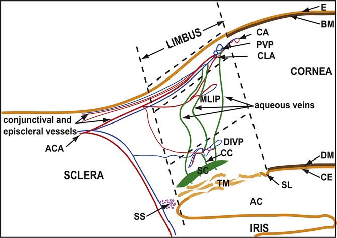
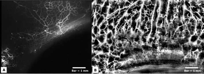
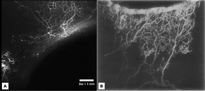
Purpose: To describe morphometric details of the human aqueous humor (AH) outflow microvasculature visualized with 360-degree virtual castings during active AH outflow in cadaver eyes and to compare these structures with corrosion casting studies.
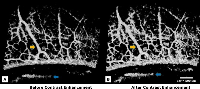
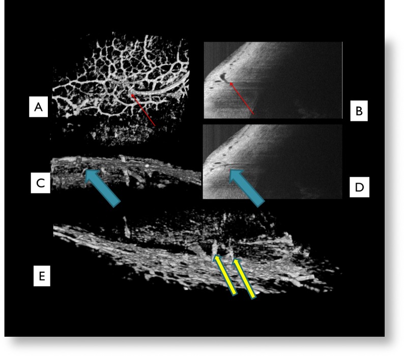
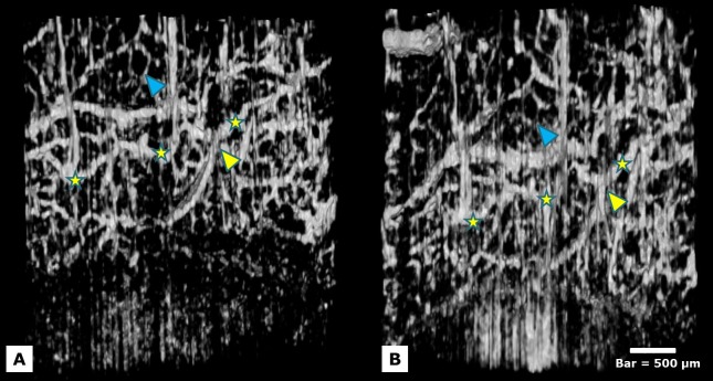
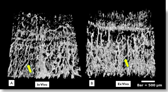
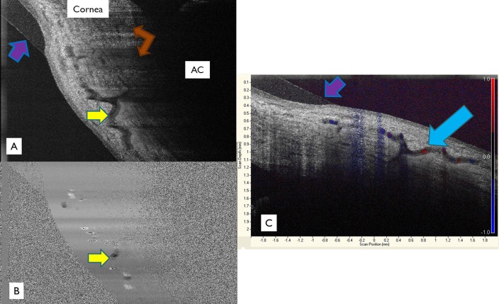
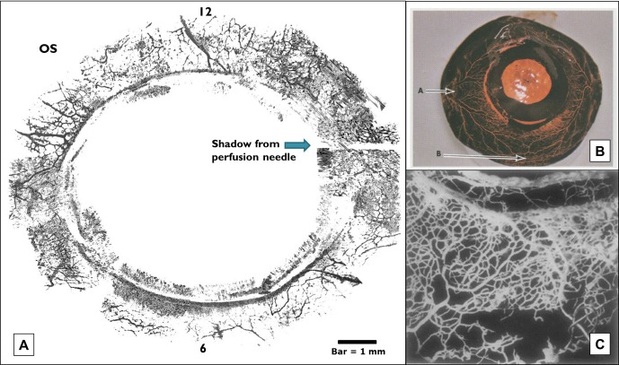
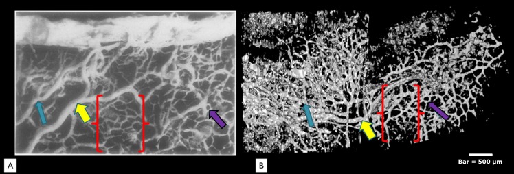
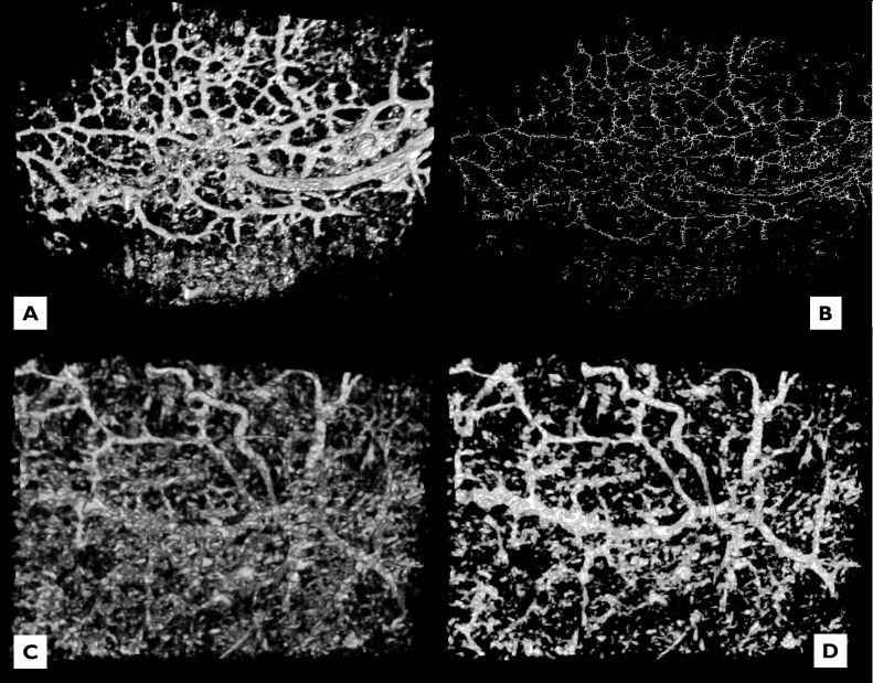
Methods: The conventional AH outflow pathways of donor eyes (n = 7) and eyes in vivo (n = 3) were imaged with spectral-domain optical coherence tomography (SD-OCT) and wide-bandwidth superluminescent diode array during active AH outflow. Digital image contrast was adjusted to isolate AH microvasculature, and images were viewed in a 3D viewer. Additional eyes (n = 3) were perfused with mock AH containing fluorescent tracer microspheres to compare microvasculature patterns.
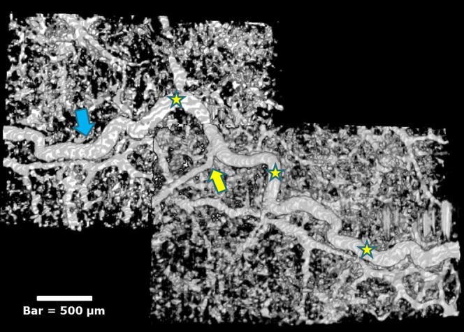
Results: Observations revealed components of the conventional outflow pathway from Schlemm's canal (SC) to the superficial intrascleral venous plexus (ISVP). The superficial ISVP in both our study and corrosion casts were composed of interconnected venules (10-50 μm) forming a hexagonal meshwork. Larger radial arcades (50-100 μm) drained the region nearest SC and converged with larger tortuous vessels (>100 μm). A 360-degree virtual casting closely approximated corrosion casting studies. Tracer studies corroborated our findings. Tracer decorated several larger vessels (50-100 μm) extending posteriorly from the limbus in both raw and contrast-enhanced fluorescence images. Smaller tracer-labeled vessels (30-40 μm) were seen branching between larger vessels and exhibited a similar hexagonal network pattern.
Conclusions: SD-OCT is capable of detailed morphometric analysis of the conventional outflow pathway in vivo or ex vivo with details comparable to corrosion casting techniques.
Conflict of interest statement
Disclosure:
Figures
References
-
- Van der Merwe EL, Kidson SH. Advances in imaging the blood and aqueous vessels of the ocular limbus. Exp Eye Res. 2010;91:118–126 - PubMed
-
- Gould DB, John SWM. Anterior segment dysgenesis and the developmental glaucomas are complex traits. Hum Mol Genet. 2002;11:1185–1193 - PubMed
-
- Gould DB, Smith RS, John SWM. Anterior segment development relevant to glaucoma. Int J Dev Biol. 2004;48:1015–1029 - PubMed
-
- Dvorak-Theobald G. Further studies on the canal of Schlemm. Am J Ophthalmol. 1955;39:65–89 - PubMed
Publication types
MeSH terms
Grants and funding
LinkOut - more resources
Full Text Sources
Other Literature Sources
