. 2012 Jun 1;287(23):19501-15.
doi: 10.1074/jbc.M112.354209. Epub 2012 Apr 12.

Unraveling unique structure and biosynthesis pathway of N-linked glycans in human fungal pathogen Cryptococcus neoformans by glycomics analysis

Affiliations

Unraveling unique structure and biosynthesis pathway of N-linked glycans in human fungal pathogen Cryptococcus neoformans by glycomics analysis

Jeong-Nam Park et al. J Biol Chem. .

Erratum in

  • J Biol Chem. 2013 Oct 4;288(40):28950

Abstract

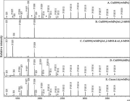
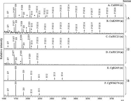
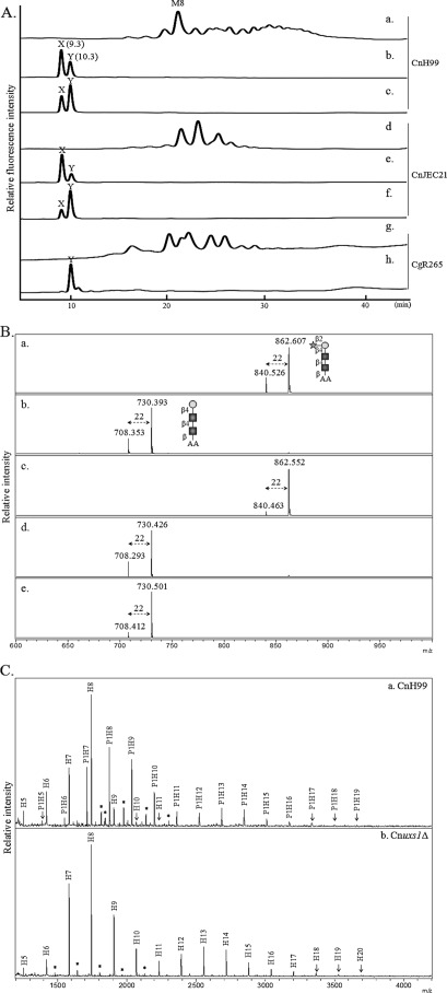
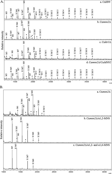
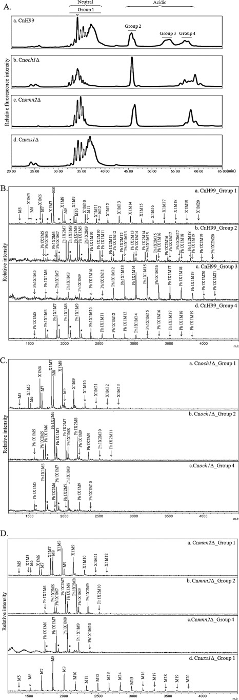
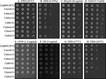
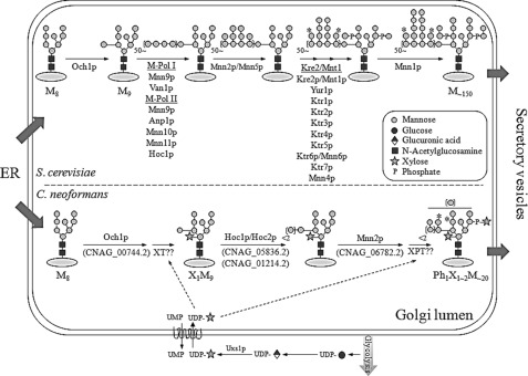
The encapsulated fungal pathogen Cryptococcus neoformans causes cryptococcosis in immunocompromised individuals. Although cell surface mannoproteins have been implicated in C. neoformans pathogenicity, the structure of N-linked glycans assembled on mannoproteins has not yet been elucidated. By analyzing oligosaccharide profiles combined with exoglycosidase treatment, we report here that C. neoformans has serotype-specific high mannose-type N-glycans with or without a β1,2-xylose residue, which is attached to the trimannosyl core of N-glycans. Interestingly, the neutral N-glycans of serotypes A and D were shown to contain a xylose residue, whereas those of serotype B appeared to be much shorter and devoid of a xylose residue. Moreover, analysis of the C. neoformans uxs1Δ mutant demonstrated that UDP-xylose is utilized as a donor sugar in N-glycan biosynthesis. We also constructed and analyzed a set of C. neoformans mutant strains lacking genes putatively assigned to the reconstructed N-glycan biosynthesis pathway. It was shown that the outer chain of N-glycan is initiated by CnOch1p with addition of an α1,6-mannose residue and then subsequently extended by CnMnn2p with multiple additions of α1,2-mannose residues. Finally, comparative analysis of acidic N-glycans from wild-type, Cnoch1Δ, Cnmnn2Δ, and Cnuxs1Δ strains strongly indicated the presence of xylose phosphate attached to mannose residues in the core and outer region of N-glycans. Our data present the first report on the unique structure and biosynthesis pathway of N-glycans in C. neoformans.


Figures

FIGURE 1.
FIGURE 1.
N-Linked oligosaccharide profiles of C. neoformans serotype A. N-Glycans of cwMPs and sMPs from the serotype A H99 strain, cultivated up to the stationary phase, were analyzed by MALDI-TOF mass spectrometry in the positive mode. A, no mannosidase treatment. B, α1,2-mannosidase (α1,2-MNS) treatment. C, α1,6-mannosidase (α1,6-MNS) treatment of α1,2-MNS-treated N-glycans. D and E, sMPs and cwMPs from the capsule-defective strain YSB42 (Cncac1Δ), respectively. The mass difference between peaks in each type of glycan is about 162 m/z, which corresponds to the mass of a single hexose residue. P, pentose; H, hexose; *, unidentified peak.
FIGURE 2.
FIGURE 2.
Comparison of N-glycan profiles between different serotypes and mating types. N-Glycans of cell wall mannoproteins from various serotypes of C. neoformans, cultivated up to the stationary phase, were analyzed by MALDI-TOF spectrometry in the positive mode. A and B, serotype A strains H99 (MATα) and KN99 (MATa), respectively; C and D, serotype D strains JEC21 (MATα) and JEC20 (MATa), respectively; and E and F, serotype B strains R265 and WM276 (both MATα), respectively. P, pentose; H, hexose; *, unidentified peak.
FIGURE 3.
FIGURE 3.
Structural analysis of xylose-containing neutral N-glycans. A, HPLC analysis. N-Glycans of C. neoformans H99 (panel a) and JEC21 (panel d) were treated serially with jack bean α-mannosidase (panels b and e) and β1,2-xylosidase (panels c and f). In the case of C. gattii R265 (panel g), the N-glycans were treated only with JBM (panel h). B, MALDI-TOF analysis. Two peaks (X and Y) from JBM-treated N-glycans of H99 (A, panel b) and JEC21 (A, panel e) as well as a single peak (Y) from JBM-treated N-glycans in R265 (A, panel h) were fractionated and analyzed by MALDI-TOF. Sodium adducts (+22) of the mass spectra (m/z) were identified as major mass peaks. Square, circle, and star symbolize N-acetylglucosamine, mannose, and xylose residues, respectively. C, N-glycan profile of Cnuxs1Δ strain. N-Glycans of cell wall mannoproteins from the C. neoformans H99 wild-type (panel a) and Cnuxs1Δ mutant (panel b) strains were analyzed by MALDI-TOF spectrometry in the positive mode. P, pentose; H, hexose; *, unidentified peak.
FIGURE 4.
FIGURE 4.
Analysis of neutral N-glycan structures and in vitro mannosyltransferase activity of the Cnoch1Δ mutant. A, N-glycan profiles of C. neoformans H99 wild-type (CnH99, panel a), Cnoch1Δ (panel b), Cnhoc1Δ (panel c), Cnhoc2Δ (panel d), and Cnoch1Δ/CnOCH1 (panel e) strains by MALDI-TOF analysis in the positive mode. B, linkage analysis of the outer region in N-glycans from the Cnoch1Δ strain without (panel a) and with treatment by α1,2- (panel b) and α1,6-MNS (panel c) treatment. X, xylose; M, mannose; *, unidentified peak. C, analysis of α1,6-mannosyltransferase activity in CnH99 and Cnoch1Δ strains. The enriched Golgi membrane fraction of CnH99 or Cnoch1Δ strain was used to analyze α1,6-mannosyltransferase activity using Man8GlcNAc2-AA as an acceptor glycan. The reaction products were then treated with α1,2-MNS. Reaction products of the membrane fractions of CnH99 and Cnoch1Δ strains (panels a and b, respectively) and the reaction products of α1,2-MNS treatment (panels c and d, respectively) were analyzed by HPLC. Squares and circles with linkage information symbolize N-acetylglucosamine and mannose, respectively.
FIGURE 5.
FIGURE 5.
Analysis of neutral N-glycan structures of Cnmnn2Δ and Cnktr3Δ mutants. A, MALDI-TOF analysis in the positive mode of N-glycans from CnH99 (panel a), Cnmnn2Δ (panel b), Cnktr3Δ (panel c), and Cnoch1Δ/CnOCH1 complemented (panel d) strains. B, linkage analysis of the outer region of N-glycans from the Cnmnn2Δ strain without (panel a) and with treatment by α1,2- (panel b) and α1,6-MNS (panel c) treatment. X, xylose; M, mannose; *, unidentified peak.
FIGURE 6.
FIGURE 6.
Acidic N-glycan analysis of C. neoformans mutant strains. A, total N-glycan profiles of C. neoformans H99 wild-type (panel a), Cnoch1Δ (panel b), Cnmnn2Δ (panel c), and Cnuxs1Δ (panel d) strains by HPLC analysis using an amine column. Triangle indicates the retention time for Man8. B, MALDI-TOF analysis in the negative reflector mode for the detection of acidic N-glycans, group 1 (panel a), group 2 (panel b), group 3 (panel c), and group 4 (panel d), released from CnH99. C, MALDI-TOF analysis in the negative reflector mode for the detection of acidic N-glycans, group 1 (panel a), group 2 (panel b), and group 4 (panel c), released from Cnoch1Δ. D, MALDI-TOF analysis in the negative reflector mode for the detection of acidic N-glycans, group 1 (panel a), group 2 (panel b), and group 4 (panel c) from Cnmnn2Δ and group 1 from Cnuxs1Δ (panel d) strains, respectively. Ph, phosphate; X, xylose; M, mannose; *, unidentified peak.
FIGURE 7.
FIGURE 7.
Phenotype analysis of C. neoformans H99 wild-type, Cnoch1Δ, Cnhoc1Δ, Cnhoc2Δ, Cnmnn2Δ, Cnktr3Δ, and Cnuxs1Δ mutant strains. Yeast cells were spotted on YPD plates only or YPD plates containing different cell wall-disturbing reagents. A, YPD at 30 °C. B, YPD with 0.05% SDS. C, YPD with 20 μg/ml hygromycin B (Hyg B). D, YPD with 7.5 mm vanadate (VAN). E, YPD with 1.5 mg/ml Calcofluor white (CFW). F, YPD with 5 mg/ml Congo red (CR). G, YPD at 37 °C. H, YPD at 39 °C. The Cnhxl1Δ mutant strain with a defect in unfolded protein response (42) was included as a positive control for a defect in cell wall integrity.
FIGURE 8.
FIGURE 8.
Proposed pathway for cryptococcal N-linked glycan biosynthesis in the Golgi. Upper and lower panels depict Golgi N-glycan biosynthetic pathways in S. cerevisiae and C. neoformans, respectively. Uncharacterized glycosyltransferases such as xylosyltransferase (XT) and xylosylphosphotransferase (XPT) are marked in the N-glycan biosynthesis pathway in C. neoformans. *, a putative site for mannose or xylose phosphorylation. Mn is a MannGlcNAc2 (n = number of mannose residue).

References

    1. Perfect J. R. (2005) Cryptococcus neoformans. A sugar-coated killer with designer genes. FEMS Immunol. Med. Microbiol. 45, 395–404 - PubMed
    1. Doering T. L. (2009) How sweet it is! Cell wall biogenesis and polysaccharide capsule formation in Cryptococcus neoformans. Annu. Rev. Microbiol. 63, 223–247 - PMC - PubMed
    1. Kwon-Chung K. J., Boekhout T., Fell J. W., Díaz M. (2002) Proposal to conserve the name Cryptococcus gattii against C. hondurianus and C. bacillisporus (Basidiomycota, Hymenomycetes, Tremellomycetidae). Taxon 51, 804–806
    1. Wozniak K. L., Levitz S. M. (2009) Isolation and purification of antigenic components of Cryptococcus. Methods Mol. Biol. 470, 71–83 - PMC - PubMed
    1. Mansour M. K., Latz E., Levitz S. M. (2006) Cryptococcus neoformans glycoantigens are captured by multiple lectin receptors and presented by dendritic cells. J. Immunol. 176, 3053–3061 - PubMed

Publication types

LinkOut - more resources