. 2012 Apr 12;74(1):49-56.
doi: 10.1016/j.neuron.2012.03.009.

Chronic pharmacological mGlu5 inhibition corrects fragile X in adult mice

Affiliations

Chronic pharmacological mGlu5 inhibition corrects fragile X in adult mice

Aubin Michalon et al. Neuron. .

Abstract

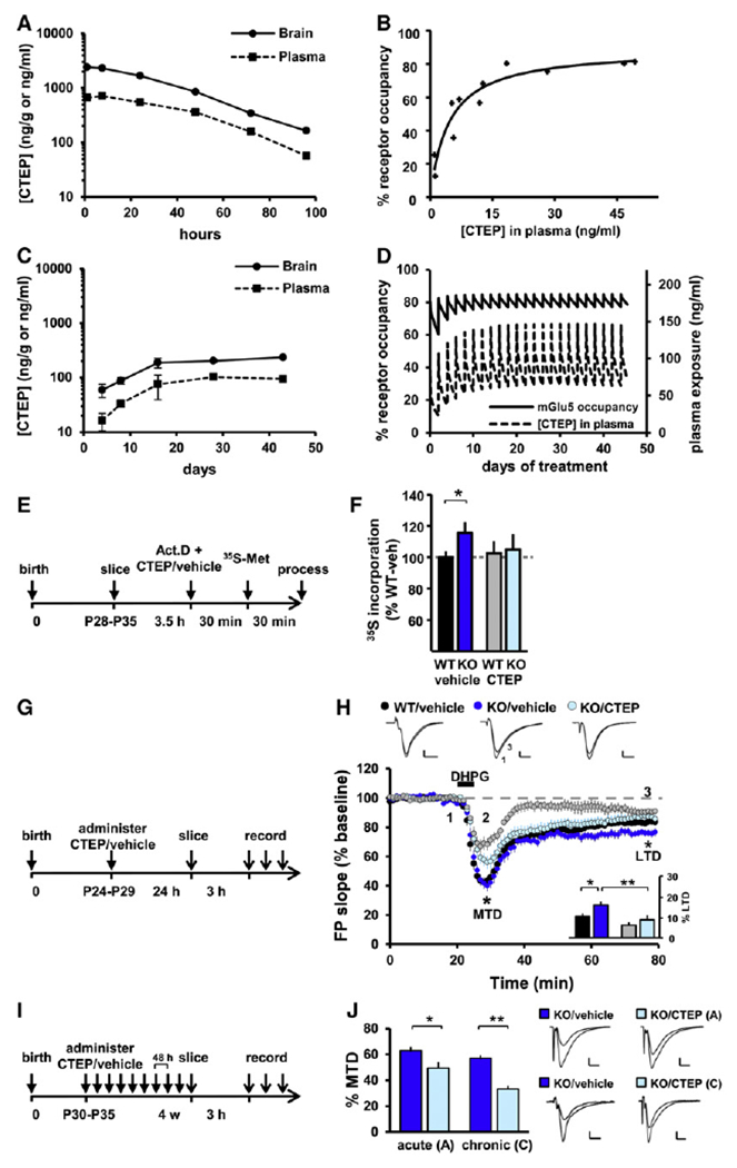
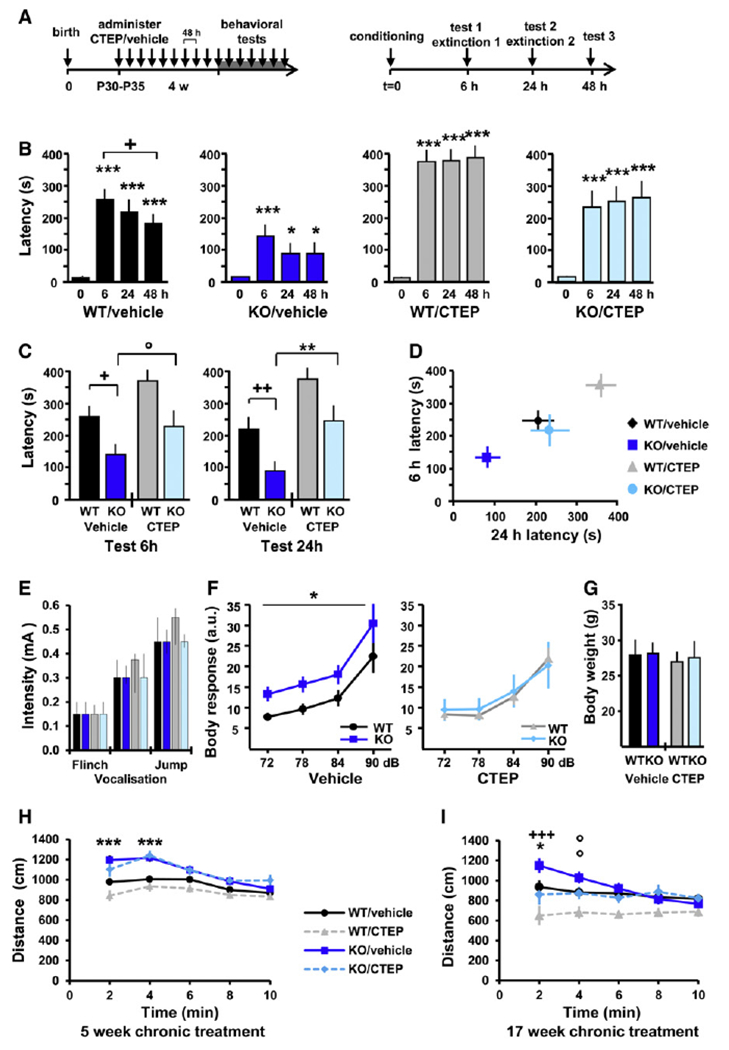
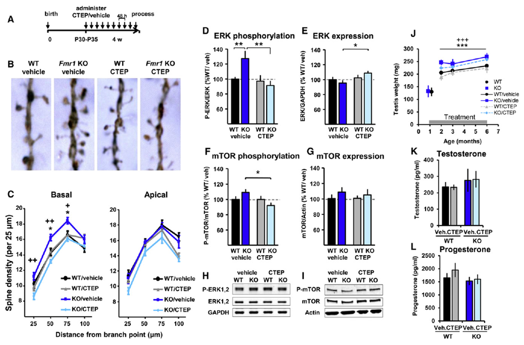
Fragile X syndrome (FXS) is the most common form of inherited intellectual disability. Previous studies have implicated mGlu5 in the pathogenesis of the disease, but a crucial unanswered question is whether pharmacological mGlu5 inhibition is able to reverse an already established FXS phenotype in mammals. Here we have used the novel, potent, and selective mGlu5 inhibitor CTEP to address this issue in the Fmr1 knockout mouse. Acute CTEP treatment corrects elevated hippocampal long-term depression, protein synthesis, and audiogenic seizures. Chronic treatment that inhibits mGlu5 within a receptor occupancy range of 81% ± 4% rescues cognitive deficits, auditory hypersensitivity, aberrant dendritic spine density, overactive ERK and mTOR signaling, and partially corrects macroorchidism. This study shows that a comprehensive phenotype correction in FXS is possible with pharmacological intervention starting in young adulthood, after development of the phenotype. It is of great interest how these findings may translate into ongoing clinical research testing mGlu5 inhibitors in FXS patients.


Figures

Figure 1.
Figure 1.. CTEP Corrects Protein Synthesis and LTD and Is Suitable for Chronic Dosing in Fmr1 KO Mice
(A) Drug exposure after a single oral dose of CTEP at 4.5 mg/kg; two mice per time point. (B) mGlu5 receptor occupancy in vivo as a function of drug exposure; 11 mice. (C) Drug-exposure monitoring during chronic dosing at 2 mg/kg per 48 hr p.o. Samples were collected 48 hr after each drug administration and thus reveal the minimal levels of drug exposure; two to four mice per time point. (D) Simulation of mGlu5 receptor occupancy during the course of 6 week chronic treatment at 2 mg/kg per 48 hr p.o. This dosing regimen achieved sustained receptor occupancy of 81% ± 4%. (E) Timeline of the protein synthesis assay measuring 35[S]-Met/Cys incorporation in hippocampus sections in vitro. (F) Protein synthesis rate in WT and Fmr1 KO slices in presence or absence of CTEP in the bath; mean ± SEM of 9–11 animals per group with two slices per animal and drug treatment; *p < 0.05. (G) Timeline of acute s.c. treatment with CTEP or vehicle 24 hr before dissection and LTD induction. (H) Gp1 mGlu LTD was enhanced in Fmr1 KO and was rescued by a single CTEP treatment. Two-way ANOVA revealed a significant effect of genotype (p < 0.05) and treatment (p < 0.01), but no interaction. Inset: post hoc tests showed a significant LTD enhancement in KO/vehicle versus WT/vehicle slices (*p < 0.05), a significant rescue by CTEP (**p < 0.01), and no significant effect of CTEP in WT slices (p = 0.14). (I) Timeline of chronic treatment schedule (2 mg/kg/48 hr p.o) beginning at the age of 4–5 weeks. (J) The maximal transient depression (MTD) induced by DHPG was significantly reduced by both acute (*p < 0.05) and chronic (**p < 0.01) CTEP treatment in KO slices, showing that the drug efficacy is maintained throughout chronic treatment. For (H) and (J), mean ± SEM of 14–18 slices per condition.
Figure 2.
Figure 2.. Pharmacological Correction of Learning and Memory Deficit and Hypersensitivity to Auditory Stimuli in Fmr1 KO Mice
(A) Timeline of chronic dosing prior to behavioral evaluation and the inhibitory avoidance and extinction (IAE) tests. (B) All experimental groups exhibited a significant increase in latency following the conditioning session (different from t = 0; *p < 0.05; **p < 0.01; ***p < 0.001), and WT/vehicle animals also showed significant memory extinction (different from t = 6 hr; +p < 0.05). (C) Comparison of latency across experimental groups at 6 and 24 hr test sessions; KO/vehicle versus WT/vehicle: *p < 0.05, **p < 0.01; KO/CTEP versus KO/vehicle: °p < 0.1, ++p < 0.01. (D) Multivariate analysis of latency at 6 versus 24 hr; the learning deficit observed in KO/vehicle mice was fully compensated by treatment, and the effect of treatment was similar in WT and Fmr1 KO mice. For (B)–(D), mean ± SEM of 15–16 mice per group. (E) Pain threshold: vehicle- and CTEP-treated Fmr1 KO and WT mice showed no differences in response to electrical foot shocks. Mean ± interquartile range, with six mice per group. (F) Sensitivity to auditory stimuli: mice were exposed to short auditory stimuli at 72 (+6), 78 (+12), 84 (+18), and 90 (+24) dB over a white background noise at 66 dB, and the whole-body startle response was recorded. Genotype effect: *p < 0.05; treatment effect: p < 0.05; mean ± SEM of 13–16 mice per group, with eight presentations of each sound intensity. (G) Body weight: there was no significant difference in body weight between the experimental groups on the day of the whole-body startle response experiment; mean ± SD of 13–16 mice per group. (H and I) Locomotor activity in the open field: Fmr1 KO mice exhibited elevated novelty-induced activity compared to WT mice at the age of 2 months (H) and 5 months (I). Correction of the hyperactivity phenotype was observed after 17 weeks (I), but not after 5 weeks (H), of chronic CTEP treatment. KO/vehicle versus WT/vehicle: °p < 0.1, *p < 0.05, ***p < 0.001; KO/CTEP versus KO/vehicle: +p < 0.05, ++p < 0.01; mean ± SEM of 16–17 (H) and 13–15 (I) mice per group.
Figure 3.
Figure 3.. Pharmacological Correction of Elevated Dendritic Spine Density and Altered Intracellular Signaling In Vivo and Partial Correction of Macroorchidism
(A) Timeline of chronic dosing for morphological and biochemical analyses. (B) Representative images of Golgi-stained neurons in the primary visual cortex; each photograph represents a 10-μm-long spine segment. (C) Spine density was increased in basal, but not apical, dendrites of KO/vehicle compared to WT/vehicle littermates (*p < 0.05) and normalized in chronically treated KO animals (KO/CTEP versus KO/vehicle: +p < 0.05, ++p < 0.01). Mean ± SEM of ten mice per group, with three dendrites on three different neurons counted per animal. (D–I) Quantification of phosphorylation and expression levels of ERK and mTOR in cortical extracts collected from chronically treated animals. (D) Elevated ERK1,2 phosphorylation (Thr202/Tyr204) in KO/vehicle mice was corrected in chronically treated KO mice (**p < 0.01). (E) Treatment also increased ERK expression levels in KO animals (*p < 0.05). (F) Similarly, mTOR phosphorylation (Ser2481) levels were significantly decreased in chronically CTEP-treated mice compared to vehicle-treated Fmr1 KO mice (*p < 0.05). (G) mTOR expression levels were not altered in Fmr1 KO mice and were not altered upon chronic treatment. For (F) and (G), mean ± SEM of 11 mice per group and triplicate measurements. (H and I) Typical western blot results. (J) Testis weight (Table S2). Adult Fmr1 KO mice presented an increased testis weight compared to WT mice (genotype effect: ***p < 0.001), which was partially corrected (~40% correction) upon chronic treatment (treatment effect: +++p < 0.001). There was no significant genotype × treatment interaction. Mean ± SD of 9–12 mice per age and per group. (K and L) Testosterone and progesterone levels were determined in the plasma of animals subjected to 17 weeks of chronic treatment. For both hormones, the levels were similar in WT and Fmr1 KO animals and were not affected by treatment. Mean ± SEM of 7–10 mice per group and duplicate measurements.

Comment in

References

    1. Banko JL, Hou L, Poulin F, Sonenberg N, and Klann E (2006). Regulation of eukaryotic initiation factor 4E by converging signaling pathways during metabotropic glutamate receptor-dependent long-term depression. J. Neurosci 26, 2167–2173. - PMC - PubMed
    1. Bear MF, Huber KM, and Warren ST (2004). The mGluR theory of fragile X mental retardation. Trends Neurosci. 27, 370–377. - PubMed
    1. Chuang SC, Zhao W, Bauchwitz R, Yan Q, Bianchi R, and Wong RK (2005). Prolonged epileptiform discharges induced by altered group I metabotropic glutamate receptor-mediated synaptic responses in hippocampal slices of a fragile X mouse model. J. Neurosci 25, 8048–8055. - PMC - PubMed
    1. Dalby KN, Morrice N, Caudwell FB, Avruch J, and Cohen P (1998). Identification of regulatory phosphorylation sites in mitogen-activated protein kinase (MAPK)-activated protein kinase-1a/p90rsk that are inducible by MAPK. J. Biol. Chem 273, 1496–1505. - PubMed
    1. Darnell JC, Van Driesche SJ, Zhang C, Hung KY, Mele A, Fraser CE, Stone EF, Chen C, Fak JJ, Chi SW, et al. (2011). FMRP stalls ribosomal translocation on mRNAs linked to synaptic function and autism. Cell 146, 247–261. - PMC - PubMed

Publication types

MeSH terms

Substances