. 2012 Apr 13:12:53.
doi: 10.1186/1471-2148-12-53.

The evolution of Dscam genes across the arthropods

Affiliations

The evolution of Dscam genes across the arthropods

Sophie A O Armitage et al. BMC Evol Biol. .

Abstract

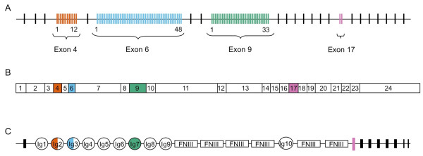
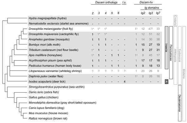
Background: One way of creating phenotypic diversity is through alternative splicing of precursor mRNAs. A gene that has evolved a hypervariable form is Down syndrome cell adhesion molecule (Dscam-hv), which in Drosophila melanogaster can produce thousands of isoforms via mutually exclusive alternative splicing. The extracellular region of this protein is encoded by three variable exon clusters, each containing multiple exon variants. The protein is vital for neuronal wiring where the extreme variability at the somatic level is required for axonal guidance, and it plays a role in immunity where the variability has been hypothesised to relate to recognition of different antigens. Dscam-hv has been found across the Pancrustacea. Additionally, three paralogous non-hypervariable Dscam-like genes have also been described for D. melanogaster. Here we took a bioinformatics approach, building profile Hidden Markov Models to search across species for putative orthologs to the Dscam genes and for hypervariable alternatively spliced exons, and inferring the phylogenetic relationships among them. Our aims were to examine whether Dscam orthologs exist outside the Bilateria, whether the origin of Dscam-hv could lie outside the Pancrustacea, when the Dscam-like orthologs arose, how many alternatively spliced exons of each exon cluster were present in the most common recent ancestor, and how these clusters evolved.

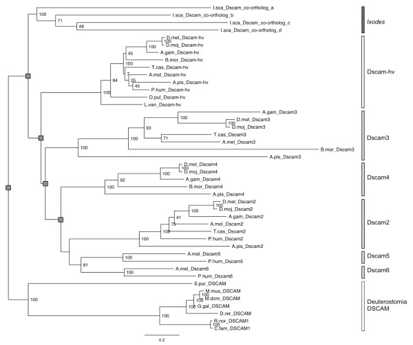
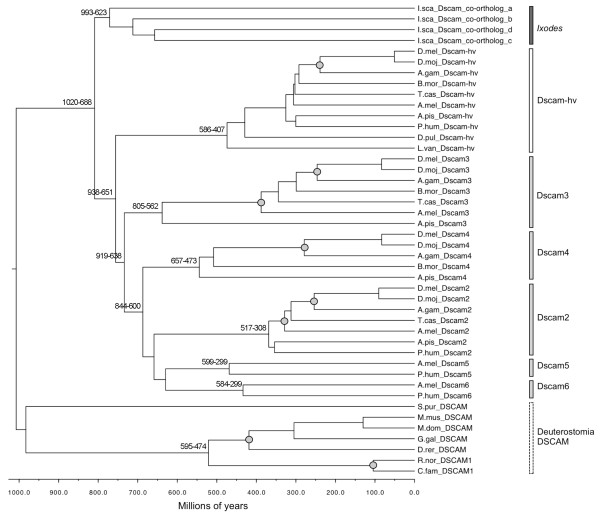
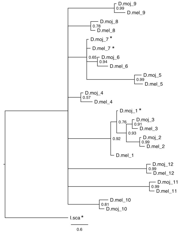
Results: Our results suggest that the origin of Dscam genes may lie after the split between the Cnidaria and the Bilateria and supports the hypothesis that Dscam-hv originated in the common ancestor of the Pancrustacea. Our phylogeny of Dscam gene family members shows six well-supported clades: five containing Dscam-like genes and one containing all the Dscam-hv genes, a seventh clade contains arachnid putative Dscam genes. Furthermore, the exon clusters appear to have experienced different evolutionary histories.

Conclusions: Dscam genes have undergone independent duplication events in the insects and in an arachnid genome, which adds to the more well-known tandem duplications that have taken place within Dscam-hv genes. Therefore, two forms of gene expansion seem to be active within this gene family. The evolutionary history of this dynamic gene family will be further unfolded as genomes of species from more disparate groups become available.


Figures

Figure 1
Figure 1
(A) Dscam-hv genomic DNA for Drosophila melanogaster. The gene consists of 20 constant exons (shown as black lines), mutually exclusive alternative splicing occurs for exons 4 (red lines), 6 (blue lines), 9 (green lines) and 17 (purple lines); one of 12 exon 4 alternatives, one of 48 exon 6 alternatives, one of 33 exon 9 alternatives and one of two exon 17 alternatives are present in each mRNA. This enables the vast number of 12 × 48 × 33 × 2 = 38,016 potential splice variants. (B) Dscam-hv mRNA. Constant exons are shown as white boxes. Exons that undergo mutually exclusive alternative splicing follow the same colour scheme as for the genomic structure. Endodomain exons 19 and 23 can be contained or lacking [8], which increases the number of potential isoforms to 4 × 38,016 = 152,064. (C) Dscam-hv protein structure for D. melanogaster. The alternatively spliced exons encode the N-terminal half of Ig2 (exon 4 in Drosophila); the N-terminal half of Ig3 (exon 6 in Drosophila), all of Ig7 (exon 9 in Drosophila), and the transmembrane domain (Exon 17 in Drosophila (figure after [6]).
Figure 2
Figure 2
Phylogenetic relationships between the species included in this study. The presence and number of Dscam orthologs and co-orthologs (I. scapularis, I.s.), Dscam-hv orthologs, and putative alternatively spliced exons within Immunoglobulins 2, 3 and 7, are indicated for each species. Grey horizontal boxes highlight the species for which our study adds some information: more specifically, black numbers show putative genes found or annotated in this study and black dashes show that we did not find Dscam orthologs/paralogs for that species. Grey numbers show the Dscam genes and Dscam-hv that were annotated or described prior to this study, although the number of Ig exon variants shown are those predicted by our HMMs. The genome of L. vannamei is not sequenced so the grey question marks indicate that we could not search for Dscam-like genes in this species. * indicates genes which were used to build HMMs to search for Dscam orthologs, and ‡ indicates genes which were used to build the Dscam-hv HMMs. § For reasoning why this is 47 and not 48, please see Additional file 2. L. vannamei alternatively spliced exon numbers are conservative estimates [26]. Vertical bars indicate the main taxa of interest in this study, where A stands for Arachnida. Species relationships from [29-35].
Figure 3
Figure 3
Maximum likelihood (RAxML) phylogeny of the Dscam/DSCAM gene family. Topology follows that of the preferred tree (Additional file 15), where grey squares indicate nodes that were fixed. Bootstrap values are shown at the non-fixed nodes. The vertical bars follow the bar colours of taxa written in black in Figure 2. The scale bar represents 0.2 substitutions per site.
Figure 4
Figure 4
Bayesian (PhyloBayes, relaxed clock) dated phylogeny of the Dscam/DSCAM gene family. 95% confidence intervals for divergence times (millions of years) are shown next to the key nodes. The x-axis shows the time scale in millions of years. The topology of the main clades follows that of the preferred tree (Figure 3 & Additional file 15), with the addition of putative gene relationships within clades being fixed to follow species relationships shown in Figure 2. Nodes used for fossil calibrations are shown with a grey circle. The vertical bars follow the bar colours of taxa written in black in Figure 2.
Figure 5
Figure 5
Bayesian (PhyloBayes) phylogeny of hypervariable Ig2 variants (exon 4) from Drosophila melanogaster and D. mojavensis. A putative Ixodes scapularis Ig2 sequence was used as the outgroup. Bootstrap values are shown at the nodes. The scale bar represents 0.6 substitutions per site. Stars indicate rogue taxa, hence their phylogenetic positions cannot be inferred with confidence.

References

    1. C. elegans Sequencing Consortium. Genome Sequence of the Nematode C. elegans: A Platform for Investigating Biology. Science. 1998;282:2012–2018. - PubMed
    1. International Human Genome Sequencing Consortium. Finishing the euchromatic sequence of the human genome. Nature. 2004;431:931–945. doi: 10.1038/nature03001. - DOI - PubMed
    1. Black DL. Mechanisms of alternative pre-messenger RNA splicing. Annu Rev Biochem. 2003;72:291–336. doi: 10.1146/annurev.biochem.72.121801.161720. - DOI - PubMed
    1. Blencowe BJ. Alternative splicing: new insights from global analyses. Cell. 2006;126(1):37–47. doi: 10.1016/j.cell.2006.06.023. - DOI - PubMed
    1. Nilsen TW, Graveley BR. Expansion of the eukaryotic proteome by alternative splicing. Nature. 2010;463:457–463. doi: 10.1038/nature08909. - DOI - PMC - PubMed

Publication types

Substances

LinkOut - more resources