. 2012 May 10;55(9):4489-500.
doi: 10.1021/jm3003697. Epub 2012 Apr 23.

Methyl effects on protein-ligand binding

Affiliations

Methyl effects on protein-ligand binding

Cheryl S Leung et al. J Med Chem. .

Abstract

The effects of addition of a methyl group to a lead compound on biological activity are examined. A literature analysis of >2000 cases reveals that an activity boost of a factor of 10 or more is found with an 8% frequency, and a 100-fold boost is a 1 in 200 event. Four cases in the latter category are analyzed in depth to elucidate any unusual aspects of the protein-ligand binding, distribution of water molecules, and changes in conformational energetics. The analyses include Monte Carlo/free-energy perturbation (MC/FEP) calculations for methyl replacements in inhibitor series for p38α MAP kinase, ACK1, PTP1B, and thrombin. Methyl substitutions ortho to an aryl ring can be particularly effective at improving activity by inducing a propitious conformational change. The greatest improvements in activity arise from coupling the conformational gain with the burial of the methyl group in a hydrophobic region of the protein.


Figures

Figure 1
Figure 1
Distribution of free energy changes on activity for substitutions of a hydrogen atom by a methyl group in publications in the Journal of Medicinal Chemistry and Bioorganic Medicinal Chemistry Letter during 2006–2011.
Figure 2
Figure 2
Representative structure from MC/FEP simulations of inhibitor 2 bound to p38α MAP kinase. The red spheres represent the positions of water molecules observed in the crystal structure. Water molecules in this computed image that are proximal to the crystallographic hydration sites are shown in stick rendering. The intermolecular hydrogen bonds are marked as blue dash lines. For clarity, nonpolar hydrogen atoms and part of the binding site are omitted.
Figure 3
Figure 3
Computed distributions for the biphenyl C-C-C-C dihedral angle (highlighted in red) for the unbound and bound p38α MAPK inhibitors.
Figure 4
Figure 4
Conformers of (a) 1 and (b) 2. The left structure is the lowest-energy one from the conformational search with GB/SA hydration and the right conformer is the one most similar to the bound structure. The ortho methyl substitution causes the two phenyl rings to be oriented in a more orthogonal manner.
Figure 5
Figure 5
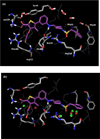
Representative configurations from the MC simulations for inhibitor 10 (a) and 11 (b) bound to ACK1. The red sphere indicates the position of a water molecule that is observed in the crystal structure of 12 complexed with ACK1. The 4-methyl group of 11 introduces steric clashes in the region of Met203, Lys158 and Glu177.
Figure 6
Figure 6
Conformers of (a) 6, (b) the C2-Me analog 7, (c) the C4-Me analog 8, (d) the C6-Me analog 9, (e) the 2,6-diMe analog 10, and (f) the 2,4,6-triMe analog 11. The left structure is the lowest-energy one from the conformational search with GB/SA hydration and the right conformer is the one most similar to the bound structure.
Figure 7
Figure 7
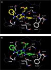
Representative structures for 14 bound to PTP1B from the MC/FEP calculations. (a) Some water molecules are illustrated in sticks that arose from the default hydration procedure. (b) The initial solvent distribution obtained using the water placement algorithm JAWS included water molecules positioned at the sites of the green spheres, which are proximal to hydration sites observed in the crystal structure for 15 (red spheres).
Figure 8
Figure 8
Representative configuration of 17 bound to thrombin from the MC/FEP calculations. Red spheres represent the positions of water molecules observed in the 2JH0 crystal structure in which 17 is also the ligand. Selected water molecules from the MC configuration are shown in stick rendering.
Figure 9
Figure 9
Left: The lowest-energy conformer of 18 (top) and the conformer distorted to the complex geometry (bottom). Right: The lowest-energy conformer of 19 (top) and the third lowest-energy conformer. Results using the OPLS/CM1A force field in the gas phase.
Figure 10
Figure 10
Renderings from the 3D7Z crystal structure of the complex of p38α MAP kinase with 2. The residues forming the hydrophobic pocket are highlighted. The space-filling rendering on the right includes the methyl group from 2 as a gold sphere.
Figure 11
Figure 11
Rendering from the 3PRZ crystal structure for a complex of 20 (Ar = 3-pyrazolyl) with PI3 kinase. The slot-like geometry of the binding site favors planar ligands.
Figure 12
Figure 12
Computed structure of 26 bound to HIV-1 reverse transcriptase.

References

    1. Barreiro EJ, Kümmerle AE, Fraga CAM. The Methylation Effect in Medicinal Chemistry. Chem. Rev. 2011;111:5215–5246. - PubMed
    1. Jorgensen WL. Free Energy Calculations, a Breakthrough for Modeling Organic Chemistry in Solution. Acc. Chem. Res. 1989;22:184–189.
    1. Kollman P. Free Energy Calculations: Applications to Chemical and Biochemical Phenomena. Chem. Rev. 1993;93:2395–2417.
    1. Lamb ML, Jorgensen WL. Curr. Opin. Chem. Biol. 1997;1:449–457. - PubMed
    1. Simonson T, Georgios A, Karplus M. Free Energy Simulations Come of Age: Protein-Ligand Recognition. Acc. Chem. Res. 2002;35:430–437. - PubMed

Publication types

MeSH terms