. 2012 Mar;17(3):037001.
doi: 10.1117/1.JBO.17.3.037001.

Instrument for fluorescence sensing of circulating cells with diffuse light in mice in vivo

Affiliations

Instrument for fluorescence sensing of circulating cells with diffuse light in mice in vivo

Eric Zettergren et al. J Biomed Opt. 2012 Mar.

Abstract

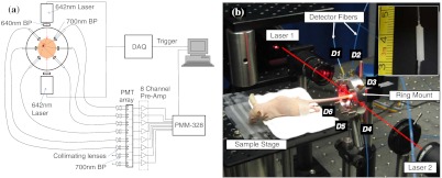
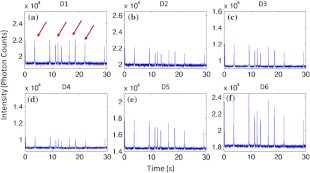
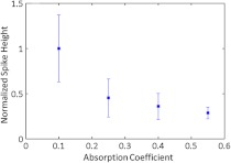
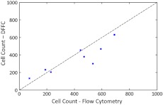
Accurate quantification of circulating cell populations in mice is important in many areas of preclinical biomedical research. Normally, this is done either by extraction and analysis of small blood samples or, more recently, by using microscopy-based in vivo fluorescence flow cytometry. We describe a new technological approach to this problem using detection of diffuse fluorescent light from relatively large blood vessels in vivo. The diffuse fluorescence flow cytometer (DFFC) uses a laser to illuminate a mouse limb and an array of optical fibers coupled to a high-sensitivity photomultiplier tube array operating in photon counting mode to detect weak fluorescence signals from cells. We first demonstrate that the DFFC instrument is capable of detecting fluorescent microspheres and Vybrant-DiD-labeled cells in a custom-made optical flow phantom with similar size, optical properties, linear flow rates, and autofluorescence as a mouse limb. We also present preliminary data demonstrating that the DFFC is capable of detecting circulating cells in nude mice in vivo. In principle, this device would allow interrogation of the whole blood volume of a mouse in minutes, with sensitivity improvement by several orders of magnitude compared to current approaches.


Figures

Fig. 1
Fig. 1
(a) A schematic of the diffuse fluorescence flow cytometry instrument, and (b) a photograph of the device during in vivo operation with a limb-mimicking optical flow phantom (inset). The position of the six detection fibers (D1 to 6) are shown.
Fig. 2
Fig. 2
Sample fluorescence signals from detector channels D1 to 6 (a–f) as microspheres passed through a limb-mimicking flow phantom at a concentration of 500  spheres/mL and a linear flow speed of 1  cm/s. Transient “spikes” (shown as arrows, panel a) were detected as each microsphere passed through the instrument field of view.
Fig. 3
Fig. 3
The amplitude of the measured fluorescent spikes as a function of the absorption coefficient of the optical flow phantom.
Fig. 4
Fig. 4
The total number of microspheres in a 1-mL sample obtained with our DFFC instrument compared to that obtained with a conventional flow cytometer. The dashed line represents the ideal 11 correspondence.
Fig. 5
Fig. 5
The FWHM of measured fluorescent spikes from fluorescent microspheres as a function of the linear flow speed through optical flow phantoms. The DFFC instrument was capable of reliably detecting microspheres over more than two orders of magnitude of flow speed.
Fig. 6
Fig. 6
Sample fluorescence signals measured from Vybrant-DiD-labeled (a) Jurkat T-lymphocyte cells (b) MM cells and (c) fluorescent microspheres through flow phantoms. Fluorescent spikes measured with the DFFC instrument were analyzed from each, and the mean and standard deviation (d) are shown. These data generally agree well with intensity analysis obtained using a commercial flow cytometer (d, inset).
Fig. 7
Fig. 7
The measured fluorescence signal from the tail of a mouse over approximately 15 min. when (a) 106 Vybrant-DiD-labeled MM cells and (b) unlabeled control cells were injected retro-orbitally. Inset: magnified sections of the curves.

References

    1. Cristofanilli M., et al. , “Circulating tumor cells, disease progression, and survival in metastatic breast cancer,” N. Engl. J. Med. 351(8), 781–791 (2004).NEJMAG10.1056/NEJMoa040766 - DOI - PubMed
    1. Smerage J. B., Hayes D. F., “The prognostic implications of circulating tumor cells in patients with breast cancer,” Cancer Invest. 26(2), 109–114 (2008).CINVD710.1080/07357900701784204 - DOI - PubMed
    1. Bauernhofer T., et al. , “Association of disease progression and poor overall survival with detection of circulating tumor cells in peripheral blood of patients with metastatic breast cancer,” Oncol. Rep. 13(2), 179–184 (2005).OCRPEW - PubMed
    1. Mocellin S., et al. , “The prognostic value of circulating tumor cells in patients with melanoma: a systematic review and meta-analysis,” Clin. Cancer Res. 12(15), 4605–4613 (2006).CCREF410.1158/1078-0432.CCR-06-0823 - DOI - PubMed
    1. Riethdorf S., et al. , “Detection of circulating tumor cells in peripheral blood of patients with metastatic breast cancer: a validation study of the CellSearch system,” Clin Cancer Res. 13(3), 920–928 (2007).CCREF410.1158/1078-0432.CCR-06-1695 - DOI - PubMed

Publication types

Substances