. 2012 Jul;130(1):205-14.e2.
doi: 10.1016/j.jaci.2012.03.002. Epub 2012 Apr 12.

Mast cell TNF receptors regulate responses to Mycoplasma pneumoniae in surfactant protein A (SP-A)-/- mice

Affiliations

Mast cell TNF receptors regulate responses to Mycoplasma pneumoniae in surfactant protein A (SP-A)-/- mice

Bethany J Hsia et al. J Allergy Clin Immunol. 2012 Jul.

Abstract

Background: Mycoplasma pneumoniae (Mp) frequently colonizes the airways of patients with chronic asthma and likely contributes to asthma exacerbations. We previously reported that mice lacking surfactant protein A (SP-A) have increased airway hyperresponsiveness (AHR) during M pneumoniae infection versus wild-type mice mediated by TNF-α. Mast cells (MCs) have been implicated in AHR in asthma models and produce and respond to TNF-α.

Objective: Determine the contribution of MC/TNF interactions to AHR in airways lacking functional SP-A during Mp infection.

Methods: Bronchoalveolar lavage fluid was collected from healthy and asthmatic subjects to examine TNF-α levels and M pneumoniae positivity. To determine how SP-A interactions with MCs regulate airway homeostasis, we generated mice lacking both SP-A and MCs (SP-A(-/-)Kit(W-sh/W-sh)) and infected them with M pneumoniae.

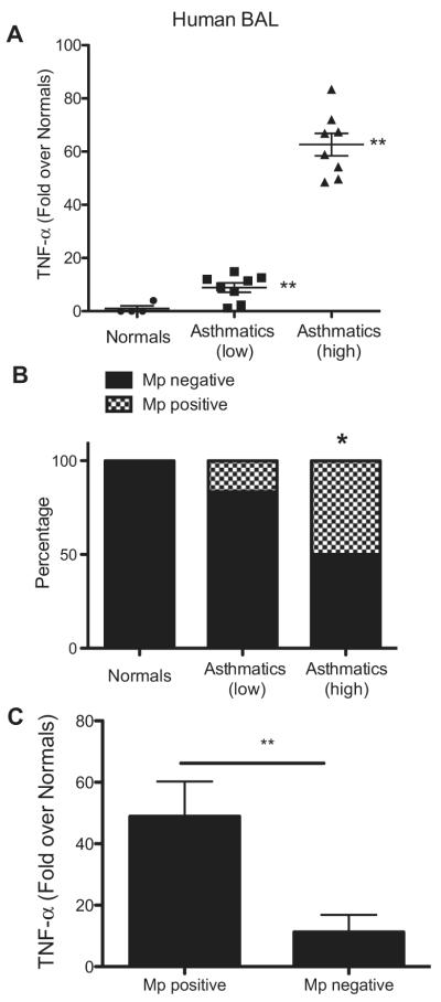
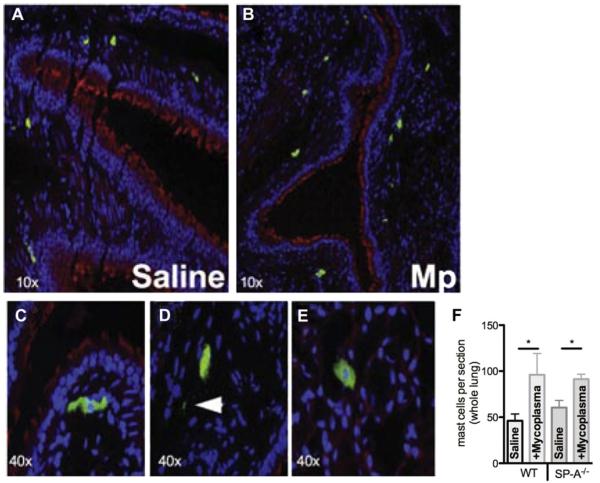
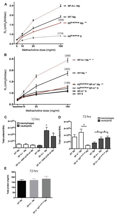
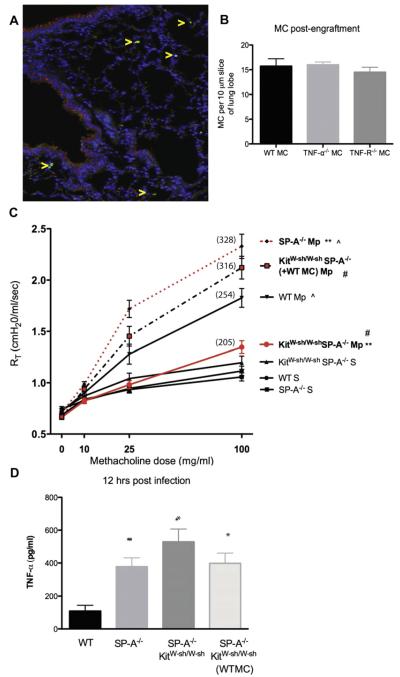
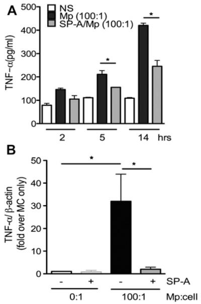
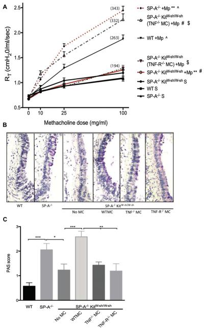
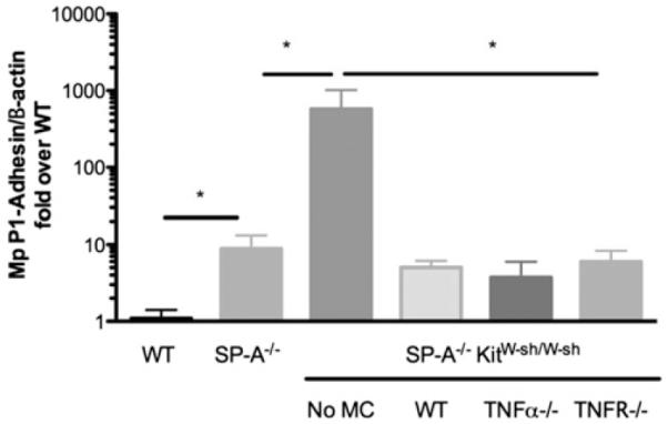
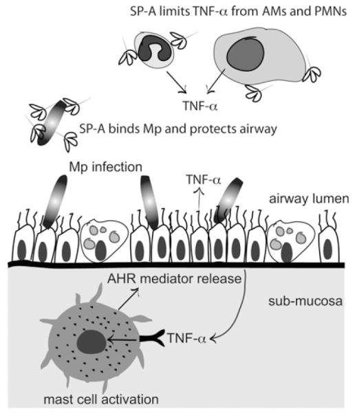
Results: Our findings indicate that high TNF-α levels correlate with M pneumoniae positivity in human asthmatic patients and that human SP-A inhibits M pneumoniae-stimulated transcription and release of TNF-α by MCs, implicating a protective role for SP-A. MC numbers increase in M pneumoniae-infected lungs, and airway reactivity is dramatically attenuated when MCs are absent. Using SP-A(-/-)Kit(W-sh/W-sh) mice engrafted with TNF-α(-/-) or TNF receptor (TNF-R)(-/-) MCs, we found that TNF-α activation of MCs through the TNF-R, but not MC-derived TNF-α, leads to augmented AHR during M pneumoniae infection when SP-A is absent. Additionally, M pneumoniae-infected SP-A(-/-)Kit(W-sh/W-sh) mice engrafted with TNF-α(-/-) or TNF-R(-/-) MCs have decreased mucus production compared with that seen in mice engrafted with wild-type MCs, whereas burden was unaffected.

Conclusion: Our data highlight a previously unappreciated but vital role for MCs as secondary responders to TNF-α during the host response to pathogen infection.


Figures

FIG 1
FIG 1
Mycoplasma pneumoniae (Mp) positivity and TNF-α levels in asthmatic samples. A, TNF-α in human BAL fluid (fold over healthy subjects). **P < .01, asthmatic patients (high) versus asthmatic patients (low) and both asthma groups versus healthy subjects. B, RT-PCR for M pneumoniae–specific P1 adhesin and dilution of samples into SP-4 broth to verify M pneumoniae positivity in subjects. *P < .05, asthmatic patients (high) versus healthy subjects and asthmatic patients (low). C, TNF values in M pneumoniae–positive and M pneumoniae–negative samples. **P < .01.
FIG 2
FIG 2
MCs in Mycoplasma pneumoniae (Mp)–infected lung tissue. Mice were instilled with saline (A) or M pneumoniae (B-E) and lungs were analyzed by means of immunohistochemistry 3 days after infection for MCs. Fig 2, C, MCs adjacent to the large airway. Fig 2, D, Extracellular MC granules (arrow). Fig 2, E, MCs in the lung parenchyma. F, Total number of tissue MCs assessed in WT versus SP-A−/− lungs (n = 5 each). *P < .05.
FIG 3
FIG 3
Contribution of MCs to AHR and inflammation. A and B, Three days after Mycoplasma pneumoniae (Mp), AHR to methacholine challenge was measured by using the Flexivent system. Mean percentage over baseline for highest methacholine dose is shown in parentheses. **P < .01 and ^P < .01, 3 independent experiments per graph. C and D, Cells in BAL fluid 12 hours (Fig 3, C) or 72 hours (Fig 3, D) after infection. *P < .05 versus WT M pneumoniae unless otherwise noted. E, Total protein in BAL fluid, n ≥ 12 mice per group.
FIG 4
FIG 4
AHR in WT bone marrow–derived MC–reconstituted infected mice. A, Engraftment of MCs (arrows) verified by means of immunohistochemistry in lung tissue 1 month after injection. B, Quantification of engraftment. C, AHR to methacholine challenge was measured by using the Flexivent system. Mean percentage over baseline for highest methacholine dose is shown in parentheses. **P < .01, #P < .01, and ^P < 0.05, 3 independent experiments per graph. D, TNF-α in BAL fluid 12 hours after infection. *P < .05 versus WT Mycoplasma pneumoniae group, n ≥ 12 mice per group. Mp, Mycoplasma pneumoniae.
FIG 5
FIG 5
TNF-α production from Mycoplasma pneumoniae (Mp)–stimulated MCs. Nonstimulated (NS) or M pneumoniae–stimulated (multiplicity of infection, 100:1) bone marrow–derived MCs with and without SP-A (40 μg/mL) were analyzed over 14 hours (A) or 1 hour (B). Supernatants were analyzed for TNF-α release by means of ELISA; cell pellets were assessed by using RT-PCR for TNF-α transcription. *P < .05. Data are representative of 3 independent experiments.
FIG 6
FIG 6
Contribution of MC TNF-R to AHR and mucus. Mice were engrafted with TNF-α−/− or TNF-R−/− bone marrow–derived MCs. A, AHR to methacholine challenge was measured by using the Flexivent system. Mean percentage over baseline for highest methacholine dose is shown in parentheses. **, ^, #, $P < .01, 3 independent experiments per graph, n ≥ 12 mice per group. B and C, PAS-stained airways in infected mice (original magnification ×10) were scored. ***P < .001, **P < .01, and *P < .05; n = 7-12 sections per group. Mp, Mycoplasma pneumoniae.
FIG 7
FIG 7
Contribution of MCs to Mycoplasma pneumoniae (Mp) clearance. M pneumoniae burden in lung tissue at day 3 was measured by using RT-PCR for the M pneumoniae–specific P1 adhesin gene relative to β-actin and is expressed as fold over WT M pneumoniae–infected mice. *P < .05, n ≥ 12 mice per group.
FIG 8
FIG 8
Protective role of SP-A against Mycoplasma pneumoniae (Mp)–induced AHR. SP-A binds M pneumoniae in the large airway and prevents overcolonization. If SP-A is dysfunctional, more M pneumoniae burden results in TNF-α overproduction from epithelial cells, alveolar macrophages(AMs), and/or neutrophils (PMNs). TNF-α encounters tissue MCs in the submucosa, which are stimulated through the TNF-R to release mediators that contribute to high levels of AHR.

Comment in

  • Findings of Research Misconduct.
    [No authors listed] [No authors listed] Fed Regist. 2019 Nov 7;84(216):60097-60098. Fed Regist. 2019. PMID: 37547121 Free PMC article. No abstract available.

References

    1. Baseman JB, Tully JG. Mycoplasmas: sophisticated, reemerging, and burdened by their notoriety. Emerg Infect Dis. 1997;3:21–32. - PMC - PubMed
    1. Krause DC, Balish MF. Cellular engineering in a minimal microbe: structure and assembly of the terminal organelle of Mycoplasma pneumoniae. Mol Microbiol. 2004;51:917–24. - PubMed
    1. Kraft M, Cassell GH, Henson JE, Watson H, Williamson J, Marmion BP, et al. Detection of Mycoplasma pneumoniae in the airways of adults with chronic asthma. Am J Respir Crit Care Med. 1998;158:998–1001. - PubMed
    1. Kraft M, Hamid Q. Mycoplasma in severe asthma. J Allergy Clin Immunol. 2006;117:1197–8. - PubMed
    1. Martin RJ, Kraft M, Chu HW, Berns EA, Cassell GH. A link between chronic asthma and chronic infection. J Allergy Clin Immunol. 2001;107:595–601. - PubMed

Publication types

MeSH terms

Substances