. 2012 May 25;36(5):742-54.
doi: 10.1016/j.immuni.2012.03.012. Epub 2012 Apr 12.

NLRP12 suppresses colon inflammation and tumorigenesis through the negative regulation of noncanonical NF-κB signaling

Affiliations

NLRP12 suppresses colon inflammation and tumorigenesis through the negative regulation of noncanonical NF-κB signaling

Irving C Allen et al. Immunity. .

Abstract

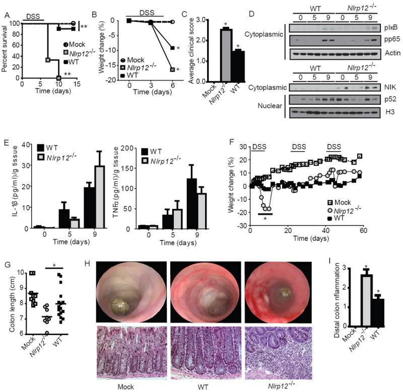
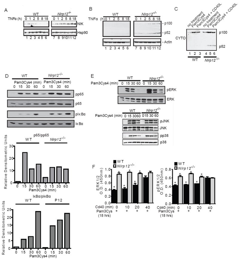
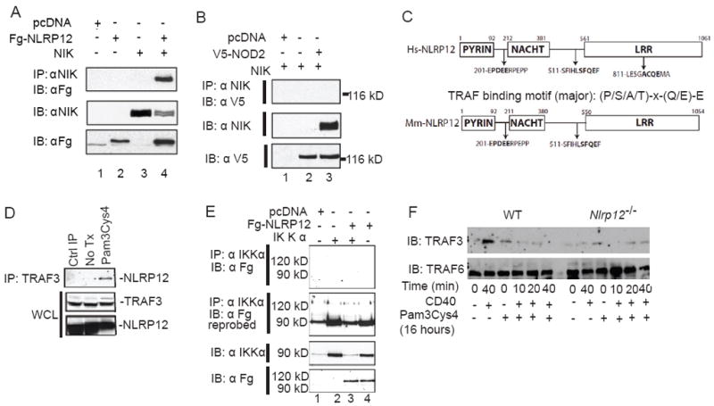
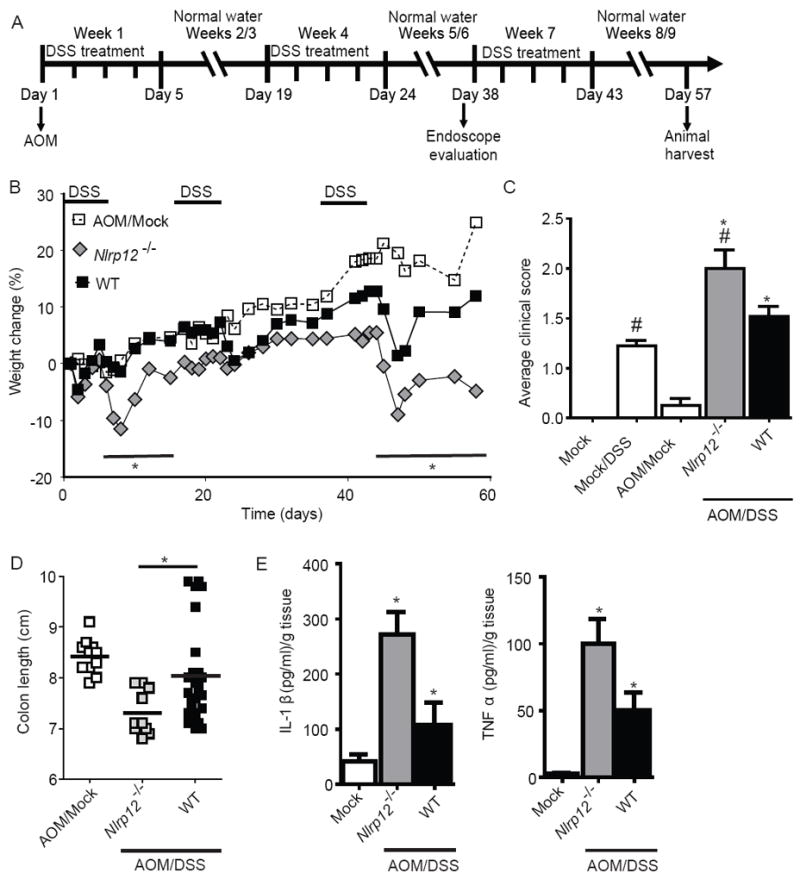
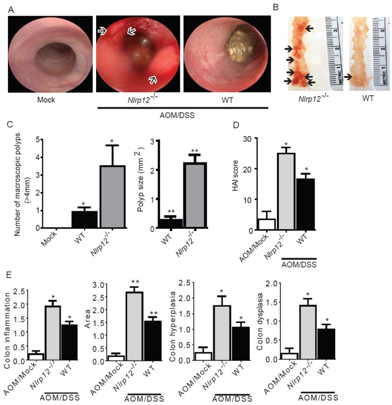
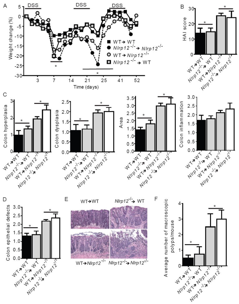
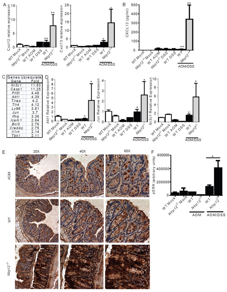
In vitro data suggest that a subgroup of NLR proteins, including NLRP12, inhibits the transcription factor NF-κB, although physiologic and disease-relevant evidence is largely missing. Dysregulated NF-κB activity is associated with colonic inflammation and cancer, and we found Nlrp12(-/-) mice were highly susceptible to colitis and colitis-associated colon cancer. Polyps isolated from Nlrp12(-/-) mice showed elevated noncanonical NF-κB activation and increased expression of target genes that were associated with cancer, including Cxcl13 and Cxcl12. NLRP12 negatively regulated ERK and AKT signaling pathways in affected tumor tissues. Both hematopoietic- and nonhematopoietic-derived NLRP12 contributed to inflammation, but the latter dominantly contributed to tumorigenesis. The noncanonical NF-κB pathway was regulated upon degradation of TRAF3 and activation of NIK. NLRP12 interacted with both NIK and TRAF3, and Nlrp12(-/-) cells have constitutively elevated NIK, p100 processing to p52 and reduced TRAF3. Thus, NLRP12 is a checkpoint of noncanonical NF-κB, inflammation, and tumorigenesis.


Figures

Figure 1
Figure 1. NLRP12 attenuates the development of experimental colitis
(A-E) Wild-type and Nlrp12-/- mice were challenged with 5% dextran sulfate sodium (DSS) for 5 days and disease progression was assessed daily. (A) Survival and (B) weight loss in Nlrp12-/- and wild-type mice. Due to increased mortality in the Nlrp12-/- mice, weight assessments were halted on Day 6. (C) Composite clinical scores reflecting weight loss, stool consistency and the presence of blood in the stool and/or rectum. (D) pIκBα, pp65 and NIK levels were evaluated in colons harvested from wild type and Nlrp12-/- mice (2 individual mice are shown per time point and all samples were run together on the same gel) prior to the initiation of acute colitis (Day 0) and 9 Days post-colitis initiation. (E) Colon amounts of IL-1β and TNFα were assessed by ELISA from organ culture supernatants. (A-E) WT mock, n = 4; DSS-treated WT, n = 10; Nlrp12-/-, n = 8. (F-I) Mice were treated with 3 rounds of 2.5% DSS for 5 days, followed by 2 weeks of recovery to assess recurring colitis. (F) Weight loss was assessed throughout the recurring DSS model. (G) Colon length from Nlrp12-/- and wild type mice. (H) Disease progression was assessed immediately prior to the third round of DSS (Day 37) via high resolution endoscopy. High resolution endoscopy was performed on 3 animals from each group. Histopathology revealed a considerable amount of crypt loss and immune cell infiltration in Nlrp12-/- mice compared to wild type mice. (I) Histopathology scoring revealed a significant increase in distal colon inflammation in the DSS treated Nlrp12-/- mice compared to the wild type animals. (F-I) WT mock, n = 9; DSS-treated WT, n = 16; Nlrp12-/-, n = 8. Data shown are representative of at least three independent experiments and depict the mean ± SEM. The symbols * and ** indicate P < 0.05 and P < 0.01, respectively, between the DSS-treated WT and Nlrp12-/- mice.
Figure 2
Figure 2. Immune cells isolated from Nlrp12-/- mice have increased non-canonical NF-κB signaling and MAPK activation
(A) Bone marrow derived dendritic cells were isolated from wild type and Nlrp12-/- mice. Following stimulation with TNFα, NIK levels steadily increased over the 18 hour time course in Nlrp12-/- cells compared to controls. (B) Increased levels of p100 cleavage to p52 were observed in Nlrp12-/- cells over duration of the TNFα challenge. (C) Cells were pretreated with Pam3Cys4 and stimulated with CD40L. Under these conditions, we detected increased p52 levels in the cytosolic fraction in cells isolated from Nlrp12-/- mice. (D) pp65 and pIκBα levels were evaluated in dendritic cells from wild type and Nlrp12-/- mice following Pam3Cys4 and CD40L stimulation. (E) Pam3Cys4 induced the phosphorylation of JNK and p38 in wild type and Nlrp12-/- cells; however, no considerable differences were consistently observed between the two genotypes. A modest increase in the phosphorylation of ERK1/2 was observed in the Nlrp12-/- cells. (F) ERK1/2 and pERK were evaluated by ELISA following Pam3Cys4 stimulation for 18 hours and CD40 stimulation over the time course shown. Primary dendritic cells from 5 independent animals were cultured and pooled to generate the ELISA data. All data shown are representative of at least three independent experiments.
Figure 3
Figure 3. NLRP12 interacts with and maintains TRAF3 levels
(A) Co-immunoprecipitation of NLRP12 and NIK following co-transfection in HEK293T cells of Fg-NLRP12 and untagged NIK. (B) NOD2 did not co-immunoprecipitate with NIK following co-transfection. (C) Bioinformatics identified multiple TRAF2/3 binding motifs in human (Hs) and murine (Mm) NLRP12. (D) NLRP12 co-immunoprecipitated with TRAF3 following 18hr Pam3Cys4 stimulation in HEK293T cells. (E) NLRP12 did not co-immunoprecipitate with IKKα following overexpression. (F) Levels of TRAF3 and TRAF6 were evaluated by immunoblot following Pam3Cys4 and CD40 stimulation in dendritic cells isolated from wild type and Nlrp12-/- mice. All data shown are representative of at least three independent experiments.
Figure 4
Figure 4. Nlrp12-/- mice are more susceptible to inflammation driven colon tumorigenesis
(A) Schematic of the inflammation driven colon tumorigenesis model. Wild type and Nlrp12-/- mice received a single injection of AOM immediately prior to the first DSS administration. The mice were then treated with 3 rounds of 2.5% DSS for 5 days, followed by 2 weeks of recovery. (B) Weight loss was monitored throughout the AOM/DSS model. (C) Composite clinical scores reflecting weight loss, stool consistency and the presence of blood in the stool and/or rectum. (D) Colons removed from AOM/DSS treated Nlrp12-/- mice were significantly truncated compared to similarly treated wild type mice. (A-D) WT mock, n = 4; Mock/DSS WT, n = 4; AOM/Mock WT, n = 11; AOM/DSS Nlrp12-/-, n = 10; AOM/DSS WT, n = 22. (E) Organ cultures were generated to determine the levels of IL-1β and TNFα in the colon. WT mock, n = 4; AOM/DSS Nlrp12-/-, n = 6; AOM/DSS WT, n = 13. For all experiments, data shown are representative of at least three independent experiments and depict the mean ± SEM. The symbols * and # indicate P < 0.05 between the AOM/DSS and DSS only-treated WT mice and the AOM/DSS treated Nlrp12-/- mice, respectively.
Figure 5
Figure 5. NLRP12 attenuates tumorigenesis during CAC
(A) Disease progression was assessed immediately prior to the third round of DSS (Day 37) via high resolution endoscopy. Excessive inflammation and hemorrhaging was observed in the Nlrp12-/- mice. Arrows denote polyp formation in the Nlrp12-/- mice. High resolution endoscopy was performed on 3 animals from each group. (B) Macroscopic polyps (arrows) were identified in the distal and mid colons harvested from Nlrp12-/- and wild type animals. (C) The number and maximal cross-sectional area of macroscopic polyps was quantified. AOM/Mock WT, n = 11; AOM/DSS Nlrp12-/-, n = 10; AOM/DSS WT, n = 22. (D) Histopathologic Activity Index (HAI) score in colons harvested following the completion of the CAC model. (E) Histopathology analysis of colon inflammation, area associated with disease, hyperplasia and dysplasia in the AOM/DSS treated Nlrp12-/- mice. WT mock, n = 5; Mock/DSS WT, n = 3; AOM/Mock WT, n = 3; AOM/DSS Nlrp12-/-, n = 5; AOM/DSS WT, n = 15. For all experiments, data shown are representative of at least three independent experiments and depict the mean ± SEM. The symbols * and ** indicate P < 0.05 and P < 0.01, respectively, between the AOM/DSS-treated WT mice and Nlrp12-/- mice.
Figure 6
Figure 6. NLRP12 mediates the development of CAC through a non-hematopoietic cellular compartment
(A) Weight change of indicated bone marrow chimera mice. (B) Composite HAI score. (C) Colon hyperplasia, dysplasia, area involved in disease and inflammation were each individually scored. (D) Nlrp12-/- mice receiving wild type or Nlrp12-/- bone marrow demonstrated a significant increase in colon area affected by disease and defects to the epithelial layer of the colon. (E) Representative histopathology emphasizing defects in the epithelial layer for each group indicated. WT→WT and Nlrp12-/-→WT shows mild to moderate surface layer thinning and tattering. WT→Nlrp12-/- and Nlrp12-/-Nlrp12-/- shows moderate to severe surface layer attenuation and erosions with multifocal detachments of epithelial islands. (F) The average number of macroscopic polyps observed in the colon. For all experiments, data shown are representative of 3 independent experiments and depict the mean ± SEM. The symbols * and # indicate P < 0.05 between the indicated groups. WT→WT, n = 5; Nlrp12-/-→WT, n = 7; WT→Nlrp12-/-, n = 7; Nlrp12-/-Nlrp12-/- n = 3.
Figure 7
Figure 7. NLRP12 attenuates noncanonical NF-κB regulated chemokines, cancer associated gene expression and specific MAPK family members during CAC
(A) Cxcl12 and Cxcl13 gene expression in colons. (B) Colon levels of CXCL13 were assessed from organ culture supernatants by ELISA. (A-B) WT mock, n = 5; Mock/DSS WT, n = 3; AOM/Mock WT, n = 3; AOM/DSS Nlrp12-/-, n = 5; AOM/DSS WT, n = 15. (C) Macroscopic polyps were removed from wild type and Nlrp12-/- mice and gene expression was assessed using a multiplex gene expression array. Data shown represent genes that were significantly up-regulated, defined as >2 fold increase, in RNA that was pooled from polyps that were microdissected from Nlrp12-/- mice compared to wild type mice. WT, n = 5; Nlrp12-/-, n = 5. (D) Gene expression for Akt1, Jun and Nr3c1 was used to verify the array data using non-pooled RNA collected from the whole colons of additional mice that were not assessed on the array. WT mock, n = 3; Nlrp12-/- mock, n = 3; WT DSS, n = 3; WT AOM, n = 3; AOM/DSS Nlrp12-/-, n = 5; AOM/DSS WT, n = 5. (E) pERK levels were evaluated by immunohistochemistry from paraffin embedded colon sections. (F) pERK levels were evaluated using semiquantitative histopathology image analysis (ImageJ). AOM/Mock WT, n = 3; AOM/DSS Nlrp12-/-, n = 6; AOM/DSS WT, n = 7.

References

    1. Adli M, Merkhofer E, Cogswell P, Baldwin AS. IKKalpha and IKKbeta each function to regulate NF-kappaB activation in the TNF-induced/canonical pathway. PLoS One. 2010 Feb 25;5(2):e9428. - PMC - PubMed
    1. Allen IC, Moore CB, Schneider M, Lei Y, Davis BK, Scull MA, Gris D, Roney KE, Zimmermann AG, Bowzard JB, et al. NLRX1 protein attenuates inflammatory responses to infection by interfering with the RIG-I-MAVS and TRAF6-NF-kappaB signaling pathways. Immunity. 2011;34:854–865. - PMC - PubMed
    1. Allen IC, TeKippe EM, Woodford RM, Uronis JM, Holl EK, Rogers AB, Herfarth HH, Jobin C, Ting JP. The NLRP3 inflammasome functions as a negative regulator of tumorigenesis during colitis-associated cancer. J Exp Med. 2010;207:1045–1056. - PMC - PubMed
    1. Annunziata CM, Davis RE, Demchenko Y, Bellamy W, Gabrea A, Zhan F, Lenz G, Hanamura I, Wright G, Xiao W, et al. Frequent engagement of the classical and alternative NF-kappaB pathways by diverse genetic abnormalities in multiple myeloma. Cancer Cell. 2007;12:115–130. - PMC - PubMed
    1. Arthur JC, Lich JD, Ye Z, Allen IC, Gris D, Wilson JE, Schneider M, Roney KE, O’Connor BP, Moore CB, et al. Cutting edge: NLRP12 controls dendritic and myeloid cell migration to affect contact hypersensitivity. J Immunol. 2010;185:4515–4519. - PMC - PubMed

Publication types

MeSH terms

Substances