. 2012 Jun;180(6):2257-67.
doi: 10.1016/j.ajpath.2012.02.010. Epub 2012 Apr 11.

Mesenchymal-specific deletion of C/EBPβ suppresses pulmonary fibrosis

Affiliations

Mesenchymal-specific deletion of C/EBPβ suppresses pulmonary fibrosis

Biao Hu et al. Am J Pathol. 2012 Jun.

Abstract

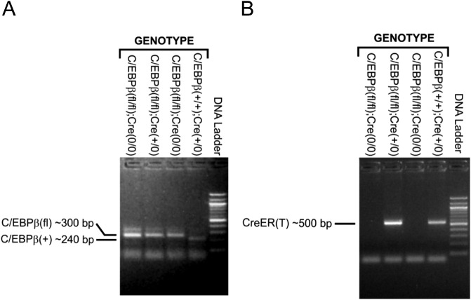
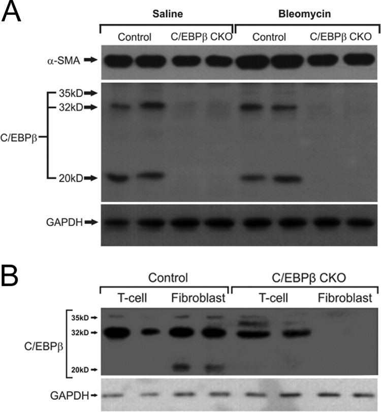
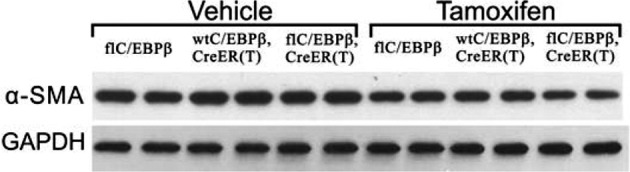
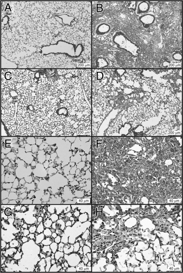
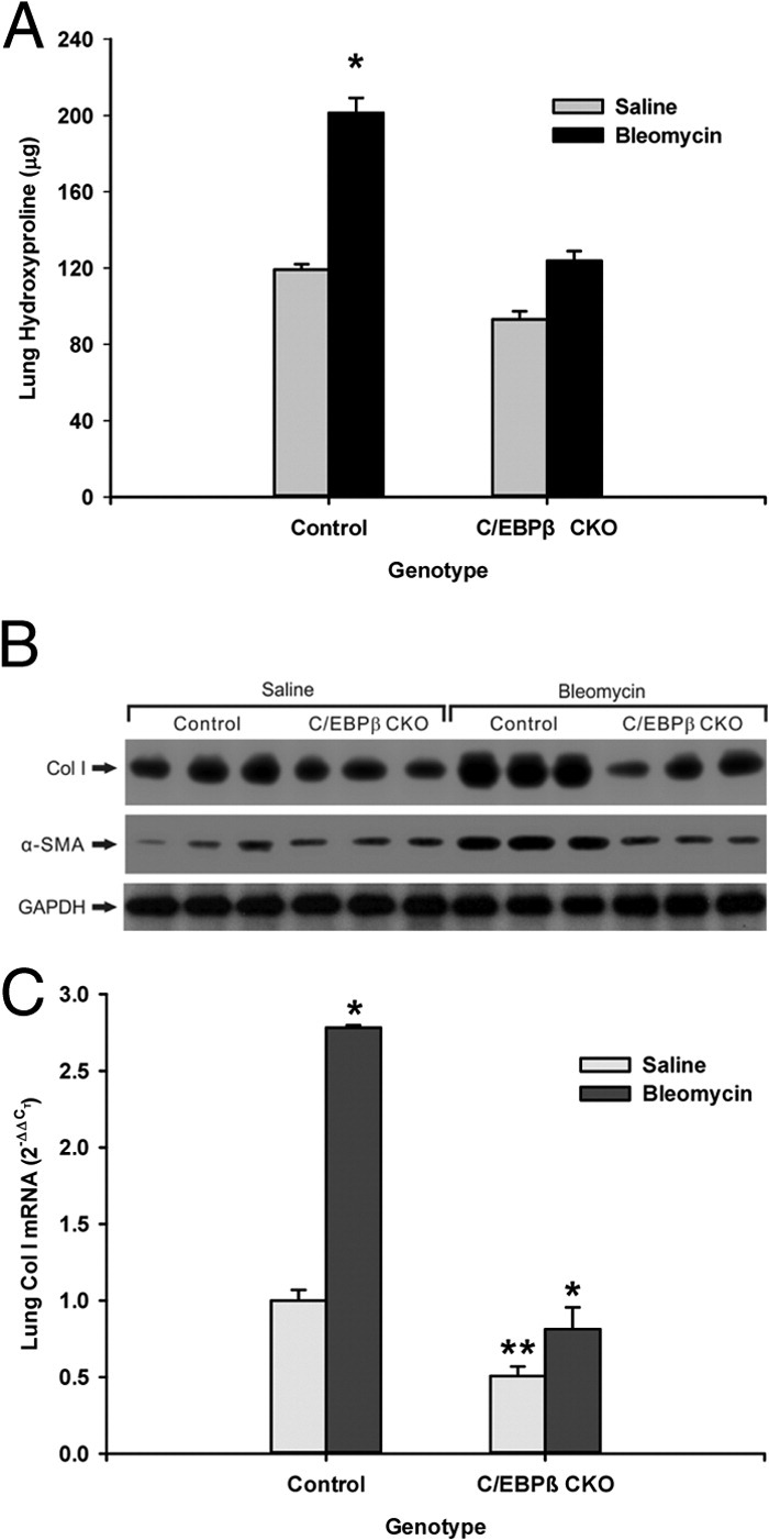
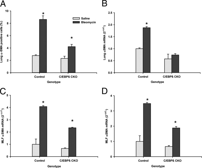
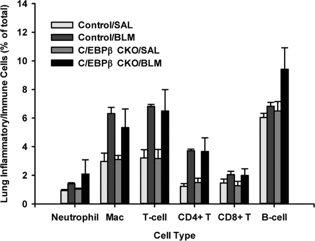
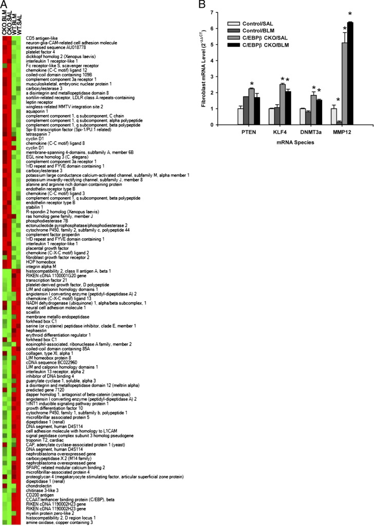
The CCAAT/enhancer-binding protein β (C/EBPβ) regulates a variety of factors and cellular responses associated with pulmonary fibrosis. To distinguish its role in the mesenchyme from that in other compartments, the effects of mesenchymal-specific deletion of C/EBPβ on pulmonary fibrosis was examined. Crossing of mice with the floxed C/EBPβ gene with α2(I) collagen enhancer-CreER(T)-bearing mice successfully generated progeny with a conditional knockout (CKO) of C/EBPβ in collagen I-expressing ("mesenchymal") cells only on treatment with tamoxifen (C/EBPβ CKO). When treated with an endotracheal bleomycin injection, C/EBPβ CKO mice showed significant attenuation of pulmonary fibrosis relative to control C/EBPβ-intact mice. C/EBPβ CKO mice also had reduced myofibroblasts in the lung. However, no significant differences in inflammatory/immune cell influx were noted in the mutant mice relative to the control mice. DNA microarray and real-time PCR analyses identified a series of myofibroblast differentiation regulators as novel target genes of C/EBPβ. Interestingly, C/EBPβ deficiency caused a marked induction of matrix metalloproteinase 12 expression, suggesting its potential role as a repressor, which could account for the noted reduction in fibrosis in the C/EBPβ-deficient mice. Thus, these findings indicate an essential role for C/EBPβ in the mesenchymal compartment in pulmonary fibrosis that is independent of its effects on inflammation or immune cell infiltration.


Figures

Figure 1
Figure 1
Mouse genotype analysis. A: PCR amplification of the C/EBPβ gene. Mouse genomic DNA was extracted and mixed with PCR primers specific for the C/EBPβ gene. The PCR products and 100-bp ladder (NEB) were separated in a 1.3% agarose gel and photographed. The ∼240-bp DNA fragment expected for the C/EBP+ allele and an ∼300-bp DNA fragment expected for the C/EBPβfl allele are indicated. B: The same genomic DNA samples were used with PCR primers specific for the CreER(T) gene. The PCR products were separated by agarose gel electrophoresis as above. The ∼500-bp amplified CreER(T) gene fragment is indicated.
Figure 2
Figure 2
Confirmation of C/EBPβ deficiency in the mesenchymal compartment. C/EBPβ CKO mice with genotype [C/EBPβfl/fl, Col1α2-Cre-ER(T)+/0] and control mice with genotype [C/EBPβ+/+, Col1α2-Cre-ER(T)+/0] were pretreated for 8 days with tamoxifen and then separated into two groups for each genotype. One group was injected endotracheally with bleomycin and the other group with PBS. Tamoxifen treatment was continued daily after the bleomycin/PBS injection until the mice were sacrificed 14 days after the bleomycin injection. A: Lung fibroblasts from these mice were analyzed for α-SMA and C/EBPβ proteins by Western blot analysis. Mice (N = 4) were tested, and a representative blot from two mice is shown. Glyceraldehyde-3-phosphate dehydrogenase (GAPDH) was used as loading control. B: Protein extracts of splenic T cells and lung fibroblasts from tamoxifen-treated C/EBPβ CKO and control mice were analyzed for C/EBPβ protein isoforms by Western blot analysis. Mice (N = 4) were tested, and a representative blot is shown with GAPDH used for loading control.
Figure 3
Figure 3
Effects of Cre and tamoxifen on α-SMA expression. Mice with the indicated genotypes were injected with tamoxifen or vehicle only as indicated. The lungs were then removed and homogenized to obtain protein extracts for analysis of α-SMA protein by Western blot analysis. Mice (N = 4) were tested, and a representative blot is shown with glyceraldehyde-3-phosphate dehydrogenase (GAPDH) as the loading control.
Figure 4
Figure 4
Effect of mesenchymal C/EBPβ deficiency on bleomycin-induced lung histopathology. Tamoxifen-treated C/EBPβ CKO mice were given endotracheal injections of either bleomycin or saline, and 21 days later lung tissue sections were stained with H&E. Representative sections from control mice treated with saline (A and E) or bleomycin (B and F) and from C/EBPβ CKO mice treated with saline (C and G) or bleomycin (D and H) are shown. Original magnification: ×10 (AD); ×40 (EH). Scale bars are included in each panel.
Figure 5
Figure 5
Effect of mesenchymal C/EBPβ deficiency on bleomycin-induced lung collagen deposition. Mice (N = 5 per group) were treated with saline or bleomycin after tamoxifen injection for 7 (real-time PCR analysis) or 21 (hydroxyproline assay and Western blot analysis) days. The lungs were harvested and analyzed for hydroxyproline content (A), type I collagen and α-SMA protein levels (B), and type I collagen mRNA levels (C). A: Results are expressed as a percentage of the respective saline control values and shown as mean ± SE (N = 5). *P < 0.05. B: Each lane represents a single animal. Glyceraldehyde-3-phosphate dehydrogenase (GAPDH) was used as a loading control after stripping the membrane. Only samples from three of the five animals are shown. Similar results were obtained from the remaining two animals (not shown). C: α1(I) procollagen mRNA was detected by real-time PCR, and the results are expressed as 2−ΔΔCT, with 18S rRNA used as the reference and the level in saline-treated wild-type mice used as calibrator. Data are shown as mean ± SE from triplicate samples. *P < 0.05 bleomycin-treated group compared with saline control; **P < 0.05 relative to the saline-treated mice with control genotype.
Figure 6
Figure 6
Effect of mesenchymal C/EBPβ deficiency on bleomycin-induced lung myofibroblast differentiation. Mice were treated with saline or bleomycin after tamoxifen injection for 7 (real-time PCR analysis) or 21 (flow cytometry and Western blot analysis) days, and the lung samples were harvested for analysis of myofibroblast differentiation. A: Single-cell suspensions were obtained from lung tissue by enzymatic digestion and analyzed for α-SMA-positive cells by flow cytometry. The results were expressed as the percentage of α-SMA-positive cells in the total lung cell suspension. B: Lung tissue RNA samples were analyzed for α-SMA mRNA by real-time PCR. C: Similar analyses of α-SMA mRNA were performed for lung fibroblasts isolated from lungs of mice treated as in A and B. D: Mouse lung fibroblasts were isolated from either C/EBPβ CKO or wild-type mice. They were treated with 4 ng/mL transforming growth factor (TGF)β for 48 hours, and the mRNAs were analyzed by real-time PCR. The mRNA results were expressed as 2−ΔΔCT, with 18S18S rRNA used as the reference and the level in saline-treated control mice (AC) or buffer-treated wild-type cells (D) used as calibrator. Data are shown as mean ± SE from triplicate samples. *P < 0.05 versus saline-treated controls (AC) and TGFβ-treated wild-type cells (D).
Figure 7
Figure 7
Effect of mesenchymal C/EBPβ deficiency on bleomycin-induced lung inflammation. Single-cell suspensions were prepared from the lungs of tamoxifen-treated C/EBPβ CKO and control mice receiving endotracheal injection of either bleomycin or saline as indicated. Seven days later, the lung samples were analyzed for the indicated inflammatory/immune cells by flow cytometry, and data were expressed as the percentage of total cells in the suspension. Data are shown as mean ± SE from five mice in each group.
Figure 8
Figure 8
Effect of mesenchymal C/EBPβ deficiency on lung fibroblast gene expression. Lung fibroblasts were isolated from C/EBPβ CKO and control mice treated with either bleomycin or saline for 7 days as indicated after the tamoxifen treatment regimen. Extracts of RNA were then subjected to DNA microarray analysis (A) or real-time PCR analysis (B). A: The expression values for each gene were calculated with a robust multiarray average method that converted the probe values into log2-transformed expression value for each gene. Genes that showed a greater than twofold difference in DNA microarray analysis between the C/EBPβ CKO cell versus control cell samples (N = 1) are shown as a heat map in order of diminishing differences toward the center (vertically). Color intensity is scaled within each row so that the highest expression value corresponds to bright red and the lowest to bright green. The sample identifiers are listed on the top of each column, and the gene names are listed to the right. B: Total RNA samples from the cells were analyzed for the indicated mRNA species by real-time PCR. The 18S rRNA was used as reference, and the saline-treated wild-type control was used as calibrator for each gene in calculation of 2−ΔΔCT. Data are shown as mean ± SE, with N = 3. *P < 0.05 versus wild-type control.

Similar articles

Cited by

References

    1. Bonner J.C. Mesenchymal cell survival in airway and interstitial pulmonary fibrosis. Fibrogenesis Tissue Repair. 2010;3:15. - PMC - PubMed
    1. Hinz B., Phan S.H., Thannickal V.J., Galli A., Bochaton-Piallat M.L., Gabbiani G. The myofibroblast: one function, multiple origins. Am J Pathol. 2007;170:1807–1816. - PMC - PubMed
    1. Phan S.H. The myofibroblast in pulmonary fibrosis. Chest. 2002;122:286S–289S. - PubMed
    1. Skalli O., Schurch W., Seemayer T., Lagace R., Montandon D., Pittet B., Gabbiani G. Myofibroblasts from diverse pathologic settings are heterogeneous in their content of actin isoforms and intermediate filament proteins. Lab Invest. 1989;60:275–285. - PubMed
    1. Darby I., Skalli O., Gabbiani G. Alpha-smooth muscle actin is transiently expressed by myofibroblasts during experimental wound healing. Lab Invest. 1990;63:21–29. - PubMed

Publication types

MeSH terms

Substances