. 2012 Jun;180(6):2536-47.
doi: 10.1016/j.ajpath.2012.03.006. Epub 2012 Apr 13.

Endometrial tumorigenesis in Pten(+/-) mice is independent of coexistence of estrogen and estrogen receptor α

Affiliations

Endometrial tumorigenesis in Pten(+/-) mice is independent of coexistence of estrogen and estrogen receptor α

Ayesha Joshi et al. Am J Pathol. 2012 Jun.

Abstract

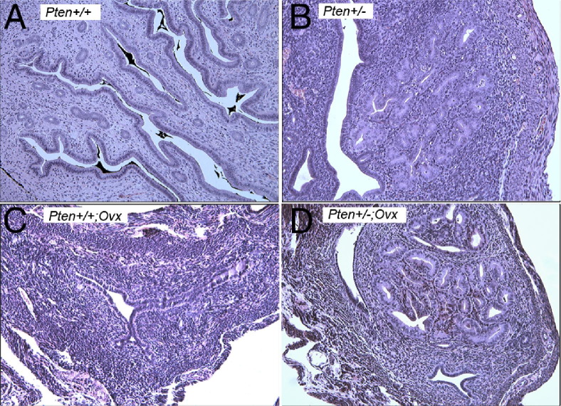
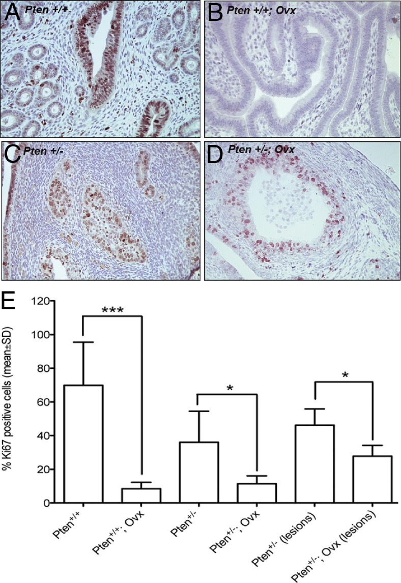
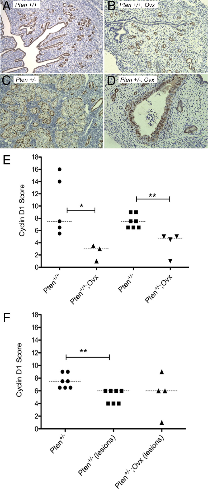
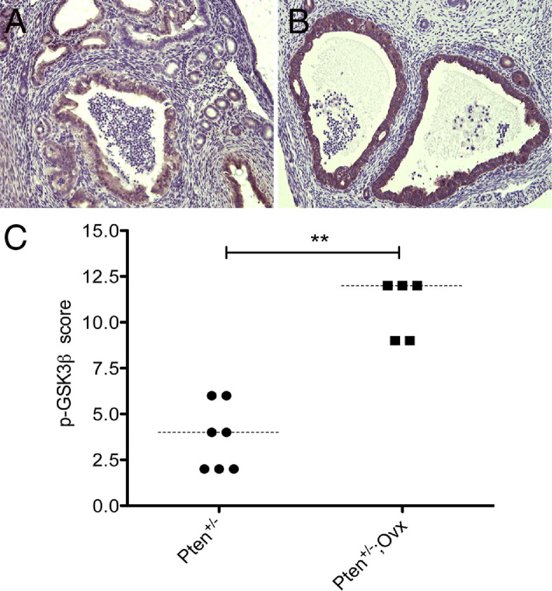
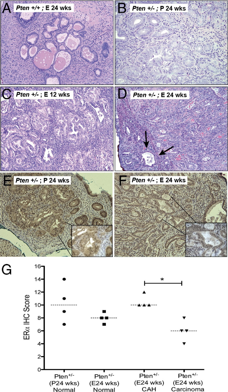
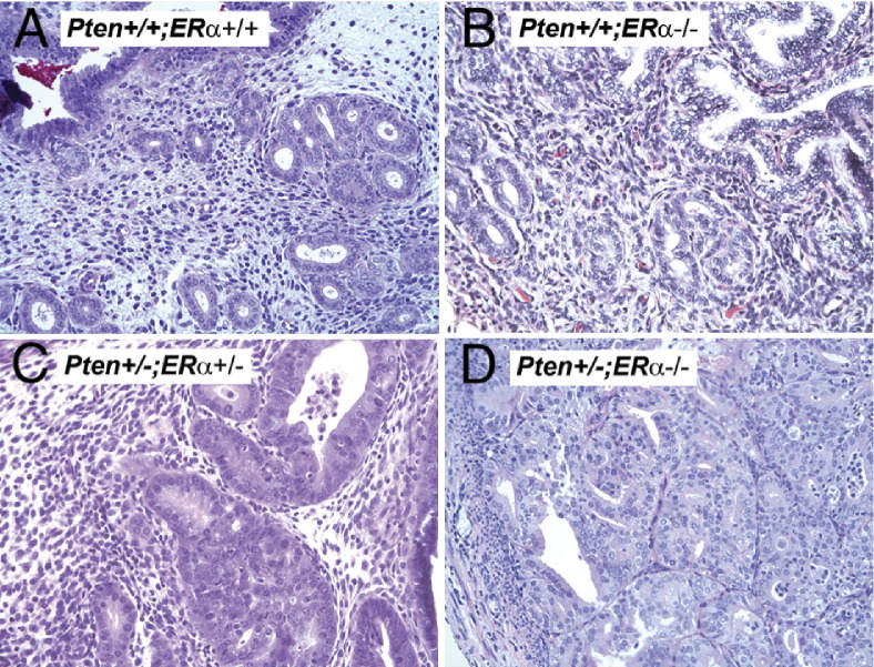
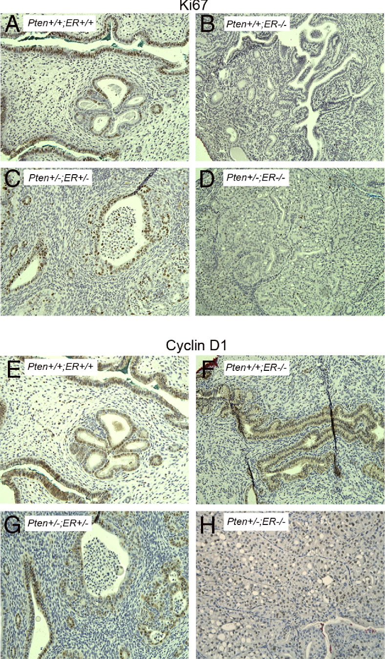
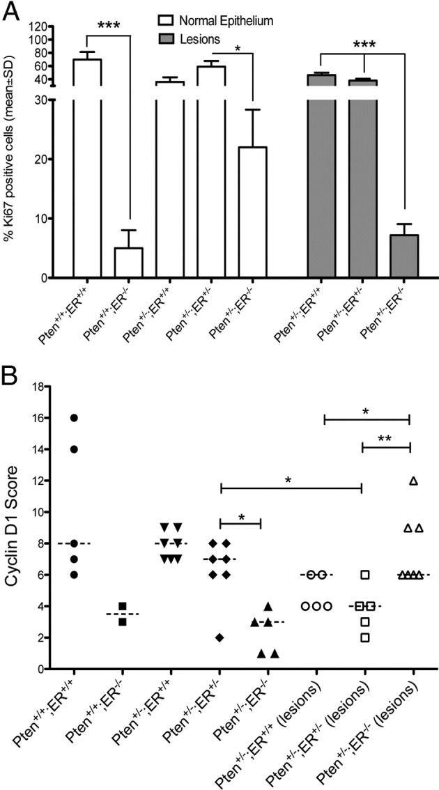
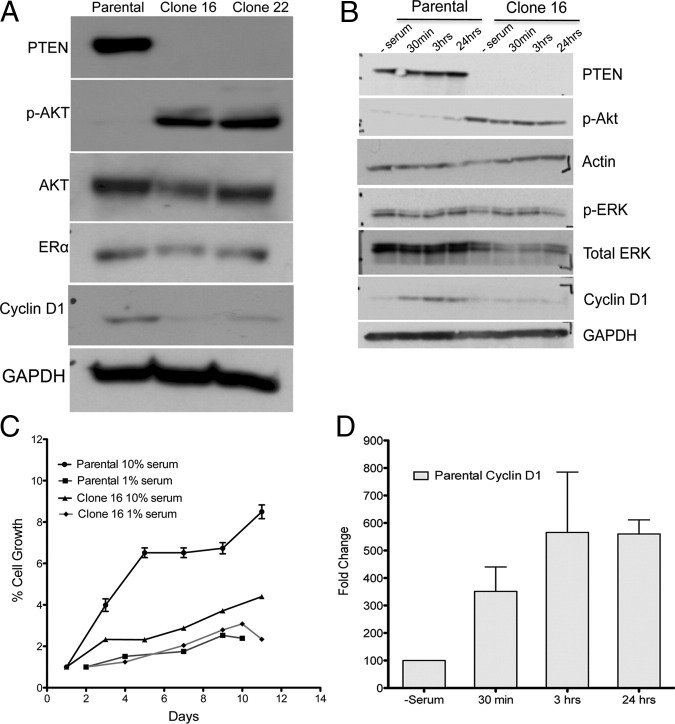
Numerous studies support the role for mutations in the phosphatase and tensin homologue (PTEN) tumor suppressor gene and unopposed estrogen stimulation in the pathogenesis of uterine endometrioid carcinoma. However, the relation between PTEN signaling and estrogen/estrogen receptor in endometrial tumorigenesis remains unresolved. We used genetically engineered mice as a model to address this relation. Mice with a single deleted Pten allele (Pten(+/-)) spontaneously develop complex atypical hyperplasia and ~20% develop endometrial cancer. To determine the effect of removing endogenous estrogen, we performed oophorectomies on Pten(+/-) mice. Although there was a reduction in the number and severity of hyperplastic lesions, the endometrial phenotype persisted, suggesting that Pten mutation, independent of estrogen, can initiate the development of complex atypical hyperplasia. To recapitulate the situation in women with unopposed estrogen, we implanted 17β-estradiol pellets in adult female Pten heterozygous mice, resulting in increased carcinoma incidence. Because studies have shown that estrogen largely acts on the endometrium via estrogen receptor ERα, we generated Pten(+/-)ERα(-/-) mice. Strikingly, 88.9% of Pten(+/-)ERα(-/-) mice developed endometrial hyperplasia/carcinoma. Furthermore, Pten(+/-)ERα(-/-) mice showed a higher incidence of in situ and invasive carcinoma, suggesting that endometrial tumorigenesis can progress in the absence of ERα. Thus, the relation between Pten alterations and estrogen signaling in the development of endometrial carcinoma is complex; the results presented herein have important implications for the treatment of endometrial hyperplasia and carcinoma in women.


Figures

Figure 1
Figure 1
H&E stained sections of Pten+/+ mice (A), Pten+/− mice (B), Pten+/+ mice that had undergone oophorectomy (Ovx) (C), and Pten+/− mice that had undergone oophorectomy (D). CAH develops in the absence of estrogen. Original magnification, ×20.
Figure 2
Figure 2
Ki-67 immunostaining on uterine sections of intact Pten+/+ mice (A), Pten+/+ mice that had undergone oophorectomy (Ovx) (B), intact Pten+/− mice (C), and Pten+/− mice that undergone oophorectomy (D). Original magnification, ×40. (E) Quantitation of Ki-67-positive cells. Oophorectomy reduced Ki-67-positive cells in the normal epithelium of Pten+/+ mice. Statistical analysis was performed with the unpaired t-test. *P values ranging between 0.01 and 0.05 were considered significant; ***P values < 0.001 were considered highly significant.
Figure 3
Figure 3
Cyclin D1 immunostaining of intact Pten+/+ mice (A), Pten+/+ mice that had undergone oophorectomy (Ovx) (B), intact Pten+/− mice (C), and Pten+/− mice that had undergone oophorectomy (D). Original magnification, ×20. (E) Quantitation of cyclin D1-positive cells. (F) Loss of Pten reduced cyclin D1 levels in CAH lesions compared with the normal epithelium. Statistical analysis was performed with the Mann–Whitney test. *P values between 0.01 and 0.05 were considered significant; **P values between 0.001 and 0.01 were considered very significant.
Figure 4
Figure 4
p-GSK3β(S9) immunostaining in CAH of intact Pten+/− mice (A) and Pten+/− mice that had undergone oophorectomy (B). Original magnification, ×20. Phosphorylation at Ser 9 was significantly up-regulated in the mice that had undergone oophorectomy (C). Statistical analysis was performed with the Mann–Whitney test. **P = 0.005.
Figure 5
Figure 5
H&E staining of uteri of CD-1 Pten+/+ mice that had undergone oophorectomy (A) treated with estradiol pellets for 24 weeks. Pten+/− mice (B) treated with placebo pellets for 24 weeks, estradiol pellets for 12 weeks (C) and 24 weeks (D). Prolonged and excess estradiol exposure increased the incidence of invasive carcinoma (arrows in D). E, estradiol; P, placebo pellets. ERα immunostaining of Pten+/− mice treated with placebo pellets (E) or estradiol pellets for 24 weeks (F). Quantitation of nuclear ERα IHC (G). Invasive carcinoma was associated with reduced nuclear ERα (Mann–Whitney test, *P = 0.02), as seen in the insets. Original magnification: ×20 (AF)l ×60 (insets of E and F).
Figure 6
Figure 6
H&E stained sections of uteri in Pten+/+;ERα+/+ (A), Pten+/+;ERα−/− (B), Pten+/−;ERα+/− (C), and Pten+/−;ERα−/− (D) mice. Carcinoma develops in the absence of ERα and invades into the myometrium. Original magnification, ×20.
Figure 7
Figure 7
Immunostaining with Ki-67 (AD) and cyclin D1 (EH) on uteri from Pten+/+;ERα+/+ (A and E), Pten+/+;ERα−/− (B and F), Pten+/−;ERα+/− (C and G), and Pten+/−;ERα−/− (D and H) mice. Invasive carcinomas in the Pten+/−;ERα−/− mice exhibit reduced number of Ki-67-positive cells but increased staining of cyclin D1. Original magnification, ×20.
Figure 8
Figure 8
Quantitation of Ki-67 (A) and Cyclin D1 (B) immunostaining depicted in Figure 7. Statistical analysis for that percentage of positive Ki-67 cells was performed with the unpaired t-test. *P values ranging between 0.01 and 0.05 were considered significant; ***P values < 0.001 were considered highly significant. For cyclin D1, Mann–Whitney test was used. *P values ranging between 0.01 and 0.05 were considered significant; **P values ranging between 0.001 and 0.01 were considered very significant.
Figure 9
Figure 9
Analysis of parental and PTEN-deleted (clones 16 and 22) Hec1A cells by immunoblot analysis (A). Deletion of PTEN activated AKT in clones 16 and 22. Both clones 16 and 22 expressed reduced cyclin D1 levels under normal growth conditions. Growth of parental cells was affected by high (10%) or low (1%) serum, whereas serum had no effect on growth of clone 16 (C). All time points for the proliferation assay were done in triplicate, and the values represent results from three independent experiments. Immunoblot analysis of PTEN, p-AKT, p-ERK, and cyclin D1 in whole-cell lysates of serum-starved cells after adding serum for indicated time periods (B) and represent the results obtained from three independent experiments. Addition of 10% serum increased cyclin D1 levels in the parental cells by at least threefold to fourfold as early as 30 minutes (D).

Similar articles

Cited by

References

    1. Di Cristofano A., Ellenson L.H. Endometrial carcinoma. Annu Rev Pathol. 2007;2:57–85. - PubMed
    1. Risinger J.I., Hayes A.K., Berchuck A., Barrett J.C. PTEN/MMAC1 mutations in endometrial cancers. Cancer Res. 1997;57:4736–4738. - PubMed
    1. Tashiro H., Blazes M.S., Wu R., Cho K.R., Bose S., Wang S.I., Li J., Parsons R., Ellenson L.H. Mutations in PTEN are frequent in endometrial carcinoma but rare in other common gynecological malignancies. Cancer Res. 1997;57:3935–3940. - PubMed
    1. Levine R.L., Cargile C.B., Blazes M.S., van Rees B., Kurman R.J., Ellenson L.H. PTEN mutations and microsatellite instability in complex atypical hyperplasia, a precursor lesion to uterine endometrioid carcinoma. Cancer Res. 1998;58:3254–3258. - PubMed
    1. Maxwell G.L., Risinger J.I., Gumbs C., Shaw H., Bentley R.C., Barrett J.C., Berchuck A., Futreal P.A. Mutation of the PTEN tumor suppressor gene in endometrial hyperplasias. Cancer Res. 1998;58:2500–2503. - PubMed

Publication types

MeSH terms