

Crowding induces live cell extrusion to maintain homeostatic cell numbers in epithelia
- PMID: 22504183
- PMCID: PMC4593481
- DOI: 10.1038/nature10999
Crowding induces live cell extrusion to maintain homeostatic cell numbers in epithelia
Abstract
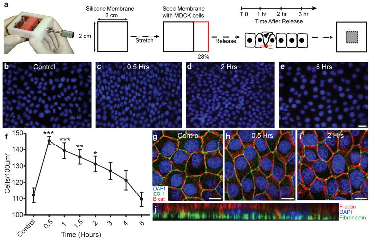
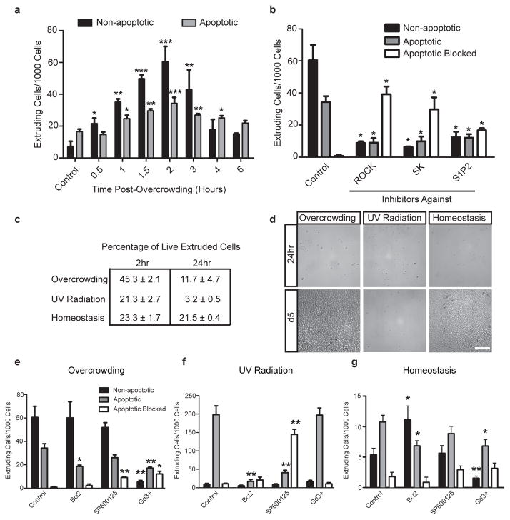
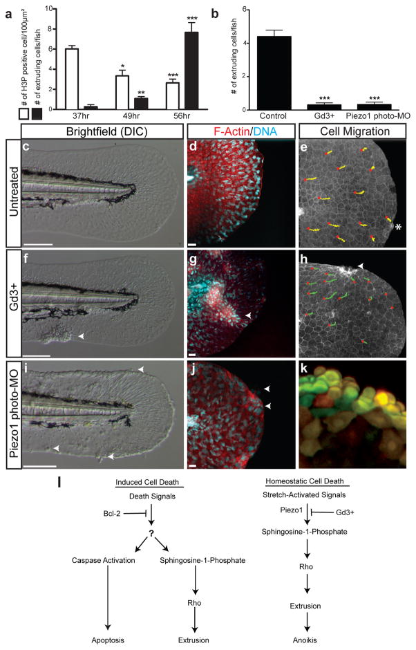
For an epithelium to provide a protective barrier, it must maintain homeostatic cell numbers by matching the number of dividing cells with the number of dying cells. Although compensatory cell division can be triggered by dying cells, it is unknown how cell death might relieve overcrowding due to proliferation. When we trigger apoptosis in epithelia, dying cells are extruded to preserve a functional barrier. Extrusion occurs by cells destined to die signalling to surrounding epithelial cells to contract an actomyosin ring that squeezes the dying cell out. However, it is not clear what drives cell death during normal homeostasis. Here we show in human, canine and zebrafish cells that overcrowding due to proliferation and migration induces extrusion of live cells to control epithelial cell numbers. Extrusion of live cells occurs at sites where the highest crowding occurs in vivo and can be induced by experimentally overcrowding monolayers in vitro. Like apoptotic cell extrusion, live cell extrusion resulting from overcrowding also requires sphingosine 1-phosphate signalling and Rho-kinase-dependent myosin contraction, but is distinguished by signalling through stretch-activated channels. Moreover, disruption of a stretch-activated channel, Piezo1, in zebrafish prevents extrusion and leads to the formation of epithelial cell masses. Our findings reveal that during homeostatic turnover, growth and division of epithelial cells on a confined substratum cause overcrowding that leads to their extrusion and consequent death owing to the loss of survival factors. These results suggest that live cell extrusion could be a tumour-suppressive mechanism that prevents the accumulation of excess epithelial cells.
Conflict of interest statement
The authors declare no competing interests.
Figures

Comment in
-
Musical chairs at the epithelium.Nat Rev Mol Cell Biol. 2012 Apr 23;13(5):279. doi: 10.1038/nrm3344. Nat Rev Mol Cell Biol. 2012. PMID: 22522715 No abstract available.
-
Feeling the squeeze: live-cell extrusion limits cell density in epithelia.Cell. 2012 May 25;149(5):965-7. doi: 10.1016/j.cell.2012.05.006. Cell. 2012. PMID: 22632965 Free PMC article.
-
Cell shedding: old questions answered.Gastroenterology. 2012 Nov;143(5):1389-1391. doi: 10.1053/j.gastro.2012.09.025. Epub 2012 Sep 20. Gastroenterology. 2012. PMID: 23000227 No abstract available.
References
-
- Ryoo HD, Gorenc T, Steller H. Apoptotic cells can induce compensatory cell proliferation through the JNK and the Wingless signaling pathways. Dev Cell. 2004;7:491–501. - PubMed
-
- Rosenblatt J, Raff MC, Cramer LP. An epithelial cell destined for apoptosis signals its neighbors to extrude it by an actin- and myosin-dependent mechanism. Curr Biol. 2001;11:1847–57. - PubMed
Publication types
MeSH terms
Substances
Grants and funding
LinkOut - more resources
Full Text Sources
Other Literature Sources
Molecular Biology Databases
