Repeatability of in vivo parafoveal cone density and spacing measurements
- PMID: 22504330
- PMCID: PMC3348369
- DOI: 10.1097/OPX.0b013e3182540562
Repeatability of in vivo parafoveal cone density and spacing measurements
Abstract
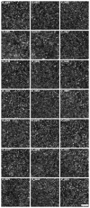
Purpose: To assess the repeatability and measurement error associated with cone density and nearest neighbor distance (NND) estimates in images of the parafoveal cone mosaic obtained with an adaptive optics scanning light ophthalmoscope (AOSLO).
Methods: Twenty-one participants with no known ocular pathology were recruited. Four retinal locations, approximately 0.65° eccentricity from the center of fixation, were imaged 10 times in randomized order with an AOSLO. Cone coordinates in each image were identified using an automated algorithm (with or without manual correction) from which cone density and NND were calculated. Owing to naturally occurring fixational instability, the 10 images recorded from a given location did not overlap entirely. We thus analyzed each image set both before and after alignment.
Results: Automated estimates of cone density on the unaligned image sets showed a coefficient of repeatability of 11,769 cones/mm(2) (17.1%). The primary reason for this variability appears to be fixational instability, as aligning the 10 images to include the exact same retinal area results in an improved repeatability of 4358 cones/mm(2) (6.4%) using completely automated cone identification software. Repeatability improved further by manually identifying cones missed by the automated algorithm, with a coefficient of repeatability of 1967 cones/mm(2) (2.7%). NND showed improved repeatability and was generally insensitive to the undersampling by the automated algorithm.
Conclusions: As our data were collected in a young, healthy population, this likely represents a best-case estimate for corresponding measurements in patients with retinal disease. Similar studies need to be carried out on other imaging systems (including those using different imaging modalities, wavefront correction technology, and/or image analysis software), as repeatability would be expected to be highly sensitive to initial image quality and the performance of cone identification algorithms. Separate studies addressing intersession repeatability and interobserver reliability are also needed.
Figures


References
-
- Liang J, Williams DR, Miller DT. Supernormal vision and high-resolution retinal imaging through adaptive optics. J Opt Soc Am (A) 1997;14:2884–92. - PubMed
-
- Roorda A, Williams DR. The arrangement of the three cone classes in the living human eye. Nature. 1999;397:520–2. - PubMed
-
- Pallikaris A, Williams DR, Hofer H. The reflectance of single cones in the living human eye. Invest Ophthalmol Vis Sci. 2003;44:4580–92. - PubMed
Publication types
MeSH terms
Grants and funding
LinkOut - more resources
Full Text Sources
Other Literature Sources
Research Materials

