. 2012 Apr 15;18(5):829-34.
doi: 10.1038/nm.2721.

A brain tumor molecular imaging strategy using a new triple-modality MRI-photoacoustic-Raman nanoparticle

Affiliations

A brain tumor molecular imaging strategy using a new triple-modality MRI-photoacoustic-Raman nanoparticle

Moritz F Kircher et al. Nat Med. .

Abstract

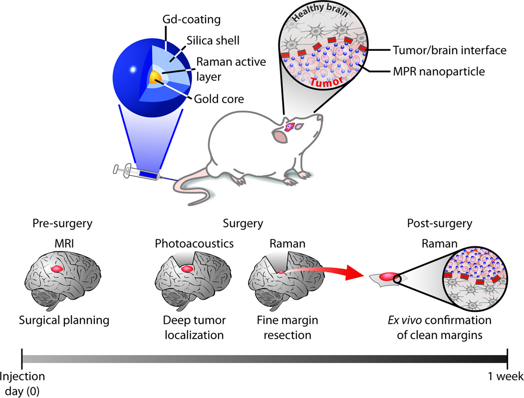
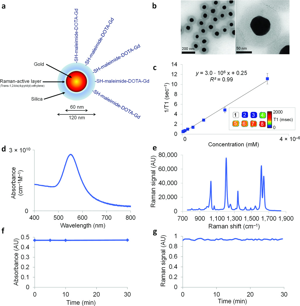
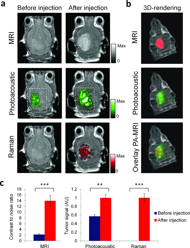
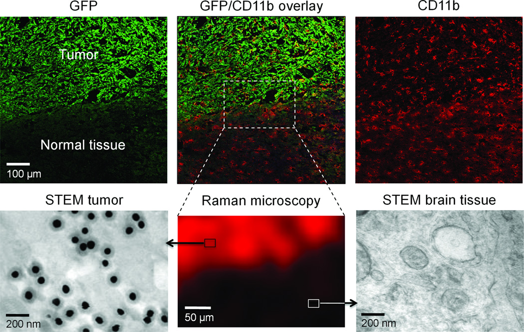
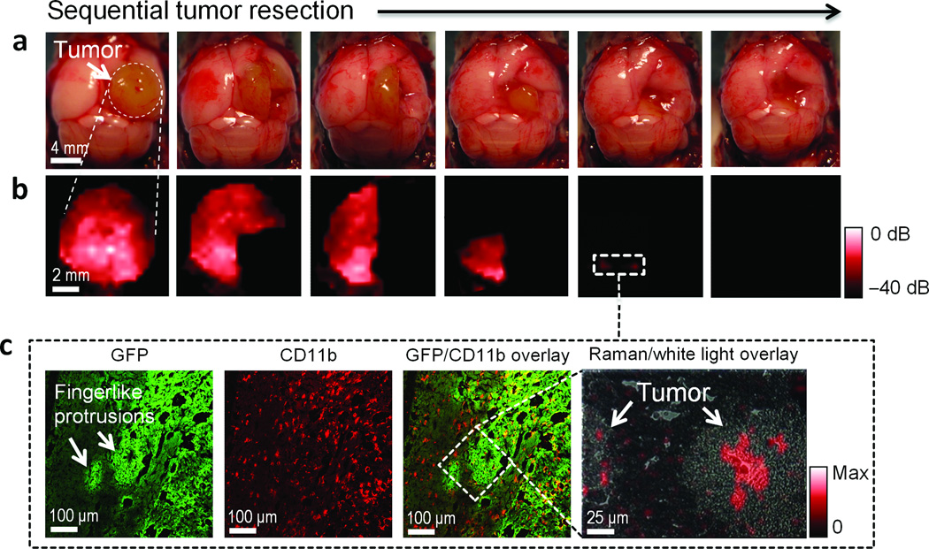
The difficulty in delineating brain tumor margins is a major obstacle in the path toward better outcomes for patients with brain tumors. Current imaging methods are often limited by inadequate sensitivity, specificity and spatial resolution. Here we show that a unique triple-modality magnetic resonance imaging-photoacoustic imaging-Raman imaging nanoparticle (termed here MPR nanoparticle) can accurately help delineate the margins of brain tumors in living mice both preoperatively and intraoperatively. The MPRs were detected by all three modalities with at least a picomolar sensitivity both in vitro and in living mice. Intravenous injection of MPRs into glioblastoma-bearing mice led to MPR accumulation and retention by the tumors, with no MPR accumulation in the surrounding healthy tissue, allowing for a noninvasive tumor delineation using all three modalities through the intact skull. Raman imaging allowed for guidance of intraoperative tumor resection, and a histological correlation validated that Raman imaging was accurately delineating the brain tumor margins. This new triple-modality-nanoparticle approach has promise for enabling more accurate brain tumor imaging and resection.


Figures

Figure 1
Figure 1. Triple-modality MPR concept
Top: MPRs are injected intravenously into a mouse bearing an orthotopic brain tumor. As the nanoparticles circulate in the blood stream, they diffuse through the disrupted blood-brain-barrier and are then sequestered and retained by the tumor. The MPRs are too large to cross the intact blood-brain-barrier and therefore cannot accumulate in healthy brain. Bottom: Concept of proposed eventual clinical use. Detectability by MRI allows pre-operative detection and surgical planning. Due to the retention of the probe, only one injection is necessary and the probe can be detected in the tumor during surgery several days later. Photoacoustic imaging with its relatively high resolution and deep tissue penetration is then able to guide bulk tumor resection intra-operatively. Raman imaging with its ultrahigh sensitivity and spatial resolution can then be used to remove residual microscopic tumor burden. Resected specimen can subsequently be examined with a Raman probe ex vivo to verify clear margins.
Figure 2
Figure 2. Characterization of the MPRs
a. Simplified diagram of the MPR. A 60 nm gold core is surrounded by a thin Raman active layer that is protected by a 30 nm silica coating. The silica coating was further functionalized with maleimide-DOTA-Gd, which was conjugated to the thiol group on the silica. b. Transmission electron microscopy images of MPRs. c. Particle relaxivity derived from T1 maps of probe dilutions in MRI phantoms. Data represent mean of two separate phantoms containing separate probe conjugations (error bars (s.e.m.) indicate batch-to-batch variation). Inset: T1 map of a MRI phantom containing MPRs at concentrations ranging from 3.2 nM (1) to 25 pM (8). d. Optical absorbance of MPRs. e. Raman spectrum of MPRs with characteristic peaks at 1,021 cm−1, 1,204 cm−1, 1,340 cm−1, 1,614 cm−1, and 1,638 cm−1. f, g. During 30 min of continuous laser irradiation, the optical absorbance (f) and the Raman signal (g) remained constant. AU, arbitrary units.
Figure 3
Figure 3. Triple-modality detection of brain tumors in living mice with MPRs
Three weeks after orthotopic inoculation, tumor-bearing mice (n = 4) were injected intravenously with MPRs (16 nM, 170 µl). Photoacoustic, Raman and MR images of the brain (skin and skull intact) were acquired before and 2 h, 3 h and 4 h after injection, respectively. a. 2D axial MRI, Photoacoustic and Raman images. The post-injection images of all three modalities demonstrated clear tumor visualization. The Photoacoustic and Raman images were co-registered with the MR image, demonstrating good co-localization between the three modalities. b. 3D-rendering of MR images with the tumor segmented (red; top); overlay of 3D Photoacoustic images (green) over MRI (middle); and overlay of MRI, segmented tumor and Photoacoustic image (bottom) showing good co-localization of the Photoacoustic signal with the tumor. c. Quantification of signal in the tumor shows significant increase in MRI, Photoacoustic and Raman signals before versus after the injection (“***” indicates P < 0.001, “**” indicates P < 0.01). Error bars represent s.e.m. AU, arbitrary units.
Figure 4
Figure 4. Histological validation
Upper row: 10 µm frozen sections from the margin of an eGFP+U87MG brain tumor were stained for eGFP (green) to visualize the tumor margins and CD11b (red) to visualize glial cells and were examined by laser scanning confocal microscopy. Bottom row: A 50 µm adjacent slice was examined by Raman microscopy to visualize the distribution of the MPRs. Note the Raman signal corresponding to the eGFP+ cells, indicating the presence of the probe in the tumor but not in the adjacent healthy tissue. The Raman signal (red) was scaled from 0 to 100 (AU). Boxes not drawn to scale. STEM images verified the presence of MPRs in the brain tissue, while no MPRs were seen in the healthy brain tissue. A three-dimensional STEM rendering of MPRs in brain tumor is provided in the Supplementary Information as Supplementary Movie.
Figure 5
Figure 5. Raman-guided intra-operative surgery using MPRs
a. Living tumor-bearing mice (n = 3) underwent craniotomy under general anesthesia. Quarters of the tumor were then sequentially removed (as illustrated in the photographs) and b. intra-operative Raman imaging was performed after each resection step, until the entire tumor had been removed by visual inspection. After the gross removal of the tumor, several small foci of Raman signal were found in the resection bed (outlined by dashed white square; some Raman images smaller than black square). Raman color scale in red from −40 to 0 dB. c. Subsequent histological analysis of sections from these foci demonstrated an infiltrative pattern of the tumor in this location, forming finger-like protrusions extending into the surrounding brain tissue. As shown in the Raman microscopy image (right), Raman signal was observed within these protrusions, indicating the selective presence of MPRs in these protrusions. Box not drawn to scale. Raman signal in linear red color scale.

Comment in

References

    1. Bucci MK, et al. Near complete surgical resection predicts a favorable outcome in pediatric patients with nonbrainstem, malignant gliomas: results from a single center in the magnetic resonance imaging era. Cancer. 2004;101:817–824. - PubMed
    1. Stupp R, et al. Changing paradigms--an update on the multidisciplinary management of malignant glioma. Oncologist. 2006;11:165–180. - PubMed
    1. Toms SA, et al. Intraoperative optical spectroscopy identifies infiltrating glioma margins with high sensitivity. Neurosurgery. 2005;57:382–391. discussion 382–391. - PubMed
    1. Orringer DA, et al. The brain tumor window model: a combined cranial window and implanted glioma model for evaluating intraoperative contrast agents. Neurosurgery. 2010;66:736–743. - PMC - PubMed
    1. Reinges MH, et al. Course of brain shift during microsurgical resection of supratentorial cerebral lesions: limits of conventional neuronavigation. Acta Neurochir (Wien) 2004;146:369–377. discussion 377. - PubMed

Publication types