. 2012 Apr 15;19(5):485-91, S1.
doi: 10.1038/nsmb.2284.

Structure of the c(10) ring of the yeast mitochondrial ATP synthase in the open conformation

Affiliations

Structure of the c(10) ring of the yeast mitochondrial ATP synthase in the open conformation

Jindrich Symersky et al. Nat Struct Mol Biol. .

Abstract

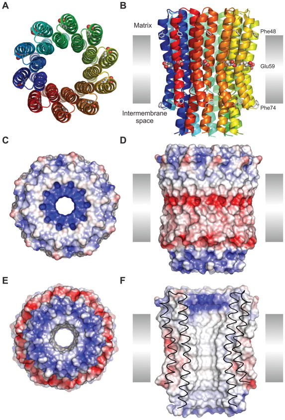
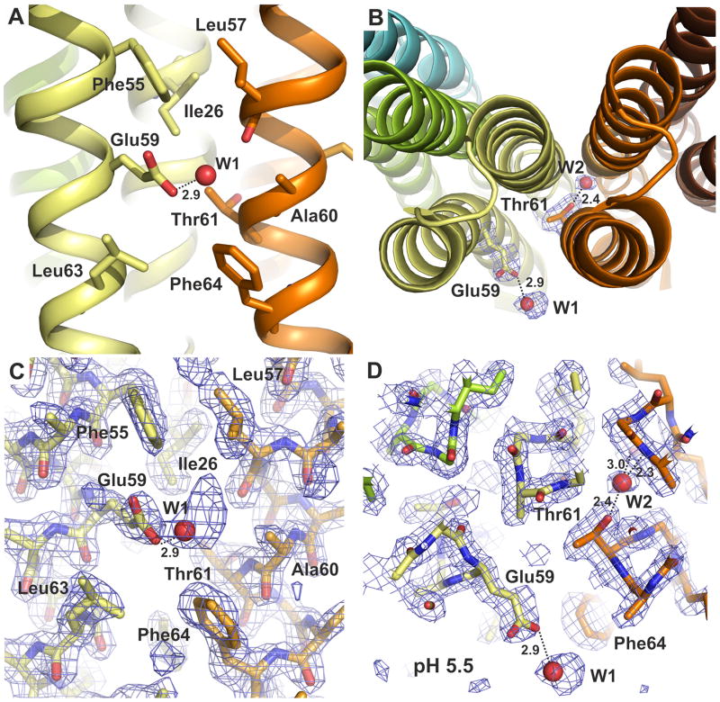
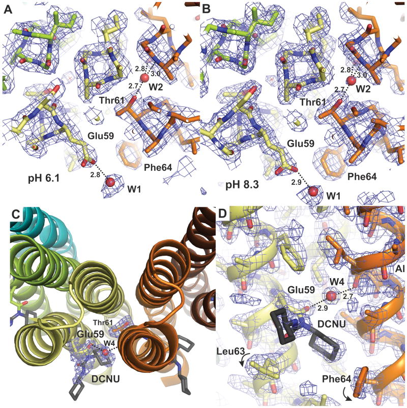
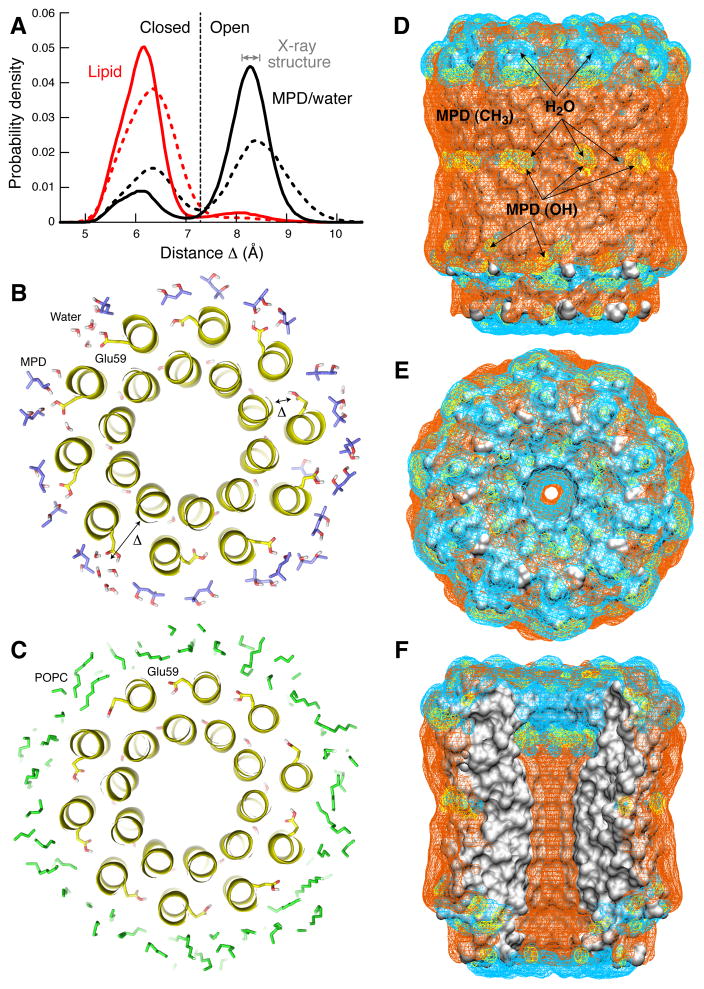
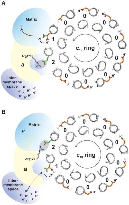
The proton pore of the F(1)F(o) ATP synthase consists of a ring of c subunits, which rotates, driven by downhill proton diffusion across the membrane. An essential carboxylate side chain in each subunit provides a proton-binding site. In all the structures of c-rings reported to date, these sites are in a closed, ion-locked state. Structures are here presented of the c(10) ring from Saccharomyces cerevisiae determined at pH 8.3, 6.1 and 5.5, at resolutions of 2.0 Å, 2.5 Å and 2.0 Å, respectively. The overall structure of this mitochondrial c-ring is similar to known homologs, except that the essential carboxylate, Glu59, adopts an open extended conformation. Molecular dynamics simulations reveal that opening of the essential carboxylate is a consequence of the amphiphilic nature of the crystallization buffer. We propose that this new structure represents the functionally open form of the c subunit, which facilitates proton loading and release.


Conflict of interest statement

Competing financial interests

The authors declare no competing financial interests.

Figures

Figure 1
Figure 1. Structure of the c10 ring from yeast ATP synthase
This figure is derived from the data obtained from the crystals grown at pH 8.3, but the results do not differ for the data sets from crystals at pH 5.5 or 6.1. A: Cartoon representation of the ring, viewed from the mitochondrial matrix, with each c-subunit in a different color. The conserved carboxyl side chain of Glu59 is shown with spheres. B: Same as A, viewed along the plane of the inner membrane (35 Å height). Phe48 and Phe74 (putative membrane limits) are represented with sticks. C: Electrostatic potential mapped on the surface of the c-ring, with positive areas in blue, negative in red, and neutral in white (color scale from −100 to 100 kBT/e). The ring is viewed from the matrix. D, E: Same as C, viewed along the plane of the membrane and from the inter-membrane space, respectively. F: Same as D, viewed in cross-section, illustrating the central hydrophobic opening of the ring.
Figure 2
Figure 2. Structure of the c10 ring proton-binding site
A: Close-up of the proton-binding site, viewed along the plane of the membrane. Residues in the vicinity are indicated, including the key carboxyl side chain, Glu59, which projects away from the binding site, and appears to form a hydrogen-bond with a solvent molecule – probably a water or the hydroxyl group of an MPD molecule. B: The proton-binding site, viewed from the matrix side of the membrane. Note that additional water molecules bridge interactions between helices, e.g. near Thr61. C, D: 2Fo-Fc electron density maps in the region of the proton-binding sites, viewed as in A, B. The maps are contoured at 1.2σ.
Figure 3
Figure 3. Protonation state of the binding site
A, B: Atomic structure of the proton binding sites in the c10 ring at pH 6.1 and 8.3, respectively, viewed as in Fig. 2D. Note the structures are essentially identical to each other, and to that at pH 5.5 (Fig. 2D). C, D: Structure of the binding site after modification with DCCD at pH 5.5, viewed either from the matrix side or along the membrane plane, respectively. The product of the reaction (DCNU) is shown in dark grey. The only significant difference in the structure of the c-ring after DCCD modification is the rotation of Leu63 and Phe64 (c.f. Fig. 2C). All 2Fo-Fc electron density maps are displayed at 1.2σ.
Figure 4
Figure 4. Simulations of the c10 ring in the lipid membrane and in the crystallization buffer
A: Probability of the open or closed conformation of the binding site, depending on whether the environment of the c-ring is the MPD:water solvent in the crystal (black) or a POPC phospholipid bilayer (red). Open and closed states are distinguished by the variable Δ, defined as the distance between the Cα atom of Ala22 and the center-of-mass of the COOH group of Glu59. Solid lines refer to simulations (100ns) in which the protein backbone is weakly constrained to the structure observed in the crystal, while side-chains are free to move. Dashed lines correspond to subsequent simulations (60ns) with no structural constraints. In the MPD:water solution, open and closed states interconvert, but the open conformation is the most likely. In the lipid membrane, the closed state is by far the most probable. B: Representative simulation snapshot of the c-ring in the MPD:water:salt solvent, viewed in cross-section; seven out of ten binding sites are open. C: Simulation snapshot of the c-ring in the lipid membrane; nine out of ten binding sites are closed. D-F: Interactions of the c10 ring structure with the crystallization buffer. All panels show 3D probability maps (mesh) for either water molecules (cyan); MPD molecules H-bonded to the protein surface via one or two of the MPD hydroxyl groups (yellow); and MPD molecules engaged in van der Waals interactions with the protein, via one or more of the MPD methyl groups (orange). The ring is viewed (D) along the plane of the membrane, (E) from the matrix side; and (F) in cross-section, illustrating the central hydrophobic opening. The MPD:water mixture provides stabilizing interactions for both polar and apolar regions on the protein surface. Note the binding sites interact either with water or with MPD hydroxyl groups.
Figure 5
Figure 5. Hypothetical rotary mechanism of proton translocation
The stator subunit-a lies adjacent to the rotor c10 ring, and provides two half channels for protons – one leading to the matrix and the other to the intermembrane space. Subunit-a also interacts with the c-ring via a conserved arginine in its fourth transmembrane helix, Arg176. When facing the membrane, the c-subunit binding sites are in the closed state and Glu59 is constitutively protonated (color orange) (State 0). The direction of rotation during ATP synthesis is counter-clockwise, viewed from the matrix (grey arrow.). A: Upon entering the subunit-a interface, a site can adopt an open conformation, and Glu59 (color green) can release a proton into the channel leading to the matrix (State 1). Glu59 in the adjacent c-subunit (in the counter-clockwise direction) is initially paired with Arg176 (State 2), but becomes free when Arg176 switches to the trailing, now deprotonated Glu59. B: The release of this interaction enables the unengaged Glu59 site to load another proton from the intermembrane space, and ultimately to re-enter the membrane.

References

    1. Kabaleeswaran V, Puri N, Walker JE, Leslie AG, Mueller DM. Novel features of the rotary catalytic mechanism revealed in the structure of yeast F1 ATPase. EMBO J. 2006;25:5433–5442. - PMC - PubMed
    1. Collinson IR, Skehel JM, Fearnley IM, Runswick MJ, Walker JE. The F1F0-ATPase complex from bovine heart mitochondria: The molar ratio of the subunits in the stalk region linking the F1 and F0 domains. Biochemistry. 1996;35:12640–12646. - PubMed
    1. Collinson IR, et al. Fo membrane domain of ATP synthase from bovine heart mitochondria: purification, subunit composition, and reconstitution with F1-ATPase. Biochemistry. 1994;33:7971–7978. - PubMed
    1. Stock D, Leslie AGW, Walker JE. Molecular architecture of the rotary motor in ATP synthase. Science. 1999;286:1700–1705. - PubMed
    1. Watt IN, Montgomery MG, Runswick MJ, Leslie AG, Walker JE. Bioenergetic cost of making an adenosine triphosphate molecule in animal mitochondria. Proc Natl Acad Sci U S A. 2010;107:16823–16827. - PMC - PubMed

Publication types

MeSH terms